 Movie
Movie Controller

[English] 日本語
 Yorodumi
- PDB-1m8v: Structure of Pyrococcus abyssii Sm Protein in Complex with a Urid... -
+ Open data

- Basic information
| Entry | Database: PDB / ID: 1m8v | |||||||||
|---|---|---|---|---|---|---|---|---|---|---|
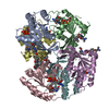
| Title | Structure of Pyrococcus abyssii Sm Protein in Complex with a Uridine Heptamer | |||||||||
 Components Components |
| |||||||||
 Keywords Keywords | RNA BINDING PROTEIN/RNA / Protein-RNA complex / Sm protein / RNA BINDING PROTEIN-RNA COMPLEX | |||||||||
| Function / homology |  Function and homology information Function and homology informationmRNA splicing, via spliceosome / ribonucleoprotein complex / RNA binding Similarity search - Function | |||||||||
| Biological species |   Pyrococcus abyssi (archaea) Pyrococcus abyssi (archaea) | |||||||||
| Method |  X-RAY DIFFRACTION / SYNCHROTRON / MOLECULAR REPLACEMENT / Resolution: 2.6 Å X-RAY DIFFRACTION / SYNCHROTRON / MOLECULAR REPLACEMENT / Resolution: 2.6 Å | |||||||||
 Authors Authors | Thore, S. / Mayer, C. / Sauter, C. / Weeks, S. / Suck, D. | |||||||||
 Citation Citation | Journal: J.Biol.Chem. / Year: 2003 Title: Crystal Structure of Pyrococcus abyssii Sm core and its Complex with RNA: Common Features of RNA-binding in Archaea and Eukarya Authors: Thore, S. / Mayer, C. / Sauter, C. / Weeks, S. / Suck, D. | |||||||||
| History |
|
- Structure visualization
| Structure viewer | Molecule: MolmilJmol/JSmol |
|---|
- Downloads & links
-Download
| PDBx/mmCIF format | 1m8v.cif.gz | 242 KB | Display | PDBx/mmCIF format |
|---|---|---|---|---|
| PDB format | pdb1m8v.ent.gz | 196.2 KB | Display | PDB format |
| PDBx/mmJSON format | 1m8v.json.gz | Tree view | PDBx/mmJSON format | |
| Others |  Other downloads Other downloads |
-Validation report
| Arichive directory | https://data.pdbj.org/pub/pdb/validation_reports/m8/1m8vftp://data.pdbj.org/pub/pdb/validation_reports/m8/1m8v | HTTPS FTP |
|---|
-Related structure data
| Related structure data | 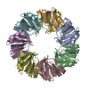 1h64SC S: Starting model for refinement C: citing same article ( |
|---|---|
| Similar structure data |


-Links
 PDBj
PDBj


- Assembly
| Deposited unit | 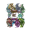
| ||||||||
|---|---|---|---|---|---|---|---|---|---|
| 1 |
| ||||||||
| Unit cell |
|
-Components
| #1: RNA chain | Mass: 2098.203 Da / Num. of mol.: 7 / Source method: obtained synthetically #2: Protein | Mass: 8630.029 Da / Num. of mol.: 14 Source method: isolated from a genetically manipulated source Source: (gene. exp.) Pyrococcus abyssi (archaea) / Organelle (production host): PLASMID / Production host:  #3: Chemical | ChemComp-CA /   Mass: 40.078 Da / Num. of mol.: 7 / Source method: obtained synthetically / Formula: Ca Mass: 40.078 Da / Num. of mol.: 7 / Source method: obtained synthetically / Formula: Ca#4: Chemical | ChemComp-U5P /   Mass: 324.181 Da / Num. of mol.: 14 / Source method: obtained synthetically / Formula: C9H13N2O9P Mass: 324.181 Da / Num. of mol.: 14 / Source method: obtained synthetically / Formula: C9H13N2O9P#5: Water | ChemComp-HOH / |  Mass: 18.015 Da / Num. of mol.: 205 / Source method: isolated from a natural source / Formula: H2O Mass: 18.015 Da / Num. of mol.: 205 / Source method: isolated from a natural source / Formula: H2OHas protein modification | N | |
|---|
-Experimental details
-Experiment
| Experiment | Method: X-RAY DIFFRACTION / Number of used crystals: 1 |
|---|
- Sample preparation
| Crystal | Density Matthews: 2.54 Å3/Da / Density % sol: 51.55 % |
|---|---|
| Crystal grow | Temperature: 277 K / Method: vapor diffusion, hanging drop / pH: 8 Details: PEG 1000, Imidazole pH8.0, Calcium acetate, VAPOR DIFFUSION, HANGING DROP, temperature 4.0K |
| Components of the solutions | Name: PEG1000 |
-Data collection
| Diffraction | Mean temperature: 100 K |
|---|---|
| Diffraction source | Source: SYNCHROTRON / Site: ESRF  / Beamline: ID29 / Wavelength: 0.915 Å / Beamline: ID29 / Wavelength: 0.915 Å |
| Detector | Type: ADSC QUANTUM 210 / Detector: CCD / Date: Nov 5, 2001 |
| Radiation | Protocol: SINGLE WAVELENGTH / Monochromatic (M) / Laue (L): M / Scattering type: x-ray |
| Radiation wavelength | Wavelength: 0.915 Å / Relative weight: 1 |
| Reflection | Resolution: 2.6→30 Å / Num. all: 40961 / Num. obs: 37089 / % possible obs: 90.5 % / Biso Wilson estimate: 28.4 Å2 / Rsym value: 0.059 / Net I/σ(I): 29.1 |
- Processing
| Software |
| |||||||||||||||||||||||||
|---|---|---|---|---|---|---|---|---|---|---|---|---|---|---|---|---|---|---|---|---|---|---|---|---|---|---|
| Refinement | Method to determine structure: MOLECULAR REPLACEMENT Starting model: PDB entry 1H64 Resolution: 2.6→30 Å / Cross valid method: THROUGHOUT
| |||||||||||||||||||||||||
| Displacement parameters | Biso mean: 38.7 Å2
| |||||||||||||||||||||||||
| Refine analyze |
| |||||||||||||||||||||||||
| Refinement step | Cycle: LAST / Resolution: 2.6→30 Å
| |||||||||||||||||||||||||
| Refine LS restraints |
|
