 Movie
Movie Controller


+ Open data

- Basic information
| Entry | Database: PDB / ID: 1auv | ||||||
|---|---|---|---|---|---|---|---|
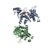
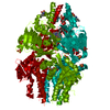
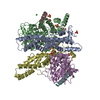
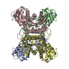
| Title | STRUCTURE OF THE C DOMAIN OF SYNAPSIN IA FROM BOVINE BRAIN | ||||||
 Components Components | SYNAPSIN IA | ||||||
 Keywords Keywords | TRANSFERASE / SYNAPSE / PHOSPHORYLATION / SYNAPSIN IA C-DOMAIN | ||||||
| Function / homology |  Function and homology information Function and homology informationsynaptic vesicle clustering / neurotransmitter secretion / synapse organization / synaptic vesicle / synaptic vesicle membrane / actin binding / Golgi apparatus / ATP binding Similarity search - Function | ||||||
| Biological species |  | ||||||
| Method |  X-RAY DIFFRACTION / SYNCHROTRON / MIR / Resolution: 2.15 Å X-RAY DIFFRACTION / SYNCHROTRON / MIR / Resolution: 2.15 Å | ||||||
 Authors Authors | Esser, L. / Wang, C. / Deisenhofer, J. | ||||||
 Citation Citation | Journal: EMBO J. / Year: 1998 Title: Synapsin I is structurally similar to ATP-utilizing enzymes. Authors: Esser, L. / Wang, C.R. / Hosaka, M. / Smagula, C.S. / Sudhof, T.C. / Deisenhofer, J. #1: Journal: Protein Sci. / Year: 1997Title: Identification, Expression, and Crystallization of the Protease-Resistant Conserved Domain of Synapsin I Authors: Wang, C.R. / Esser, L. / Smagula, C.S. / Sudhof, T.C. / Deisenhofer, J. #2: Journal: Science / Year: 1989Title: Synapsins: Mosaics of Shared and Individual Domains in a Family of Synaptic Vesicle Phosphoproteins Authors: Sudhof, T.C. / Czernik, A.J. / Kao, H.T. / Takei, K. / Johnston, P.A. / Horiuchi, A. / Kanazir, S.D. / Wagner, M.A. / Perin, M.S. / De Camilli, P. / Greengard, P. | ||||||
| History |
|
- Structure visualization
| Structure viewer | Molecule: MolmilJmol/JSmol |
|---|
- Downloads & links
-Download
| PDBx/mmCIF format | 1auv.cif.gz | 131.4 KB | Display | PDBx/mmCIF format |
|---|---|---|---|---|
| PDB format | pdb1auv.ent.gz | 102.9 KB | Display | PDB format |
| PDBx/mmJSON format | 1auv.json.gz | Tree view | PDBx/mmJSON format | |
| Others |  Other downloads Other downloads |
-Validation report
| Arichive directory | https://data.pdbj.org/pub/pdb/validation_reports/au/1auvftp://data.pdbj.org/pub/pdb/validation_reports/au/1auv | HTTPS FTP |
|---|
-Related structure data


-Links
 PDBj
PDBj


- Assembly
| Deposited unit | 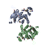
| ||||||||
|---|---|---|---|---|---|---|---|---|---|
| 1 | 
| ||||||||
| Unit cell |
| ||||||||
| Noncrystallographic symmetry (NCS) | NCS oper: (Code: given Matrix: (-0.414908, -0.346441, 0.841326), Vector: |
-Components
| #1: Protein | Mass: 35417.820 Da / Num. of mol.: 2 / Fragment: RESIDUES 110 - 420 Source method: isolated from a genetically manipulated source Source: (gene. exp.)  #2: Water | ChemComp-HOH / |  Mass: 18.015 Da / Num. of mol.: 161 / Source method: isolated from a natural source / Formula: H2O Mass: 18.015 Da / Num. of mol.: 161 / Source method: isolated from a natural source / Formula: H2OHas protein modification | Y | |
|---|
-Experimental details
-Experiment
| Experiment | Method: X-RAY DIFFRACTION / Number of used crystals: 1 |
|---|
- Sample preparation
| Crystal | Density Matthews: 2.15 Å3/Da / Density % sol: 42.83 % Description: DATA WERE COLLECTED USING THE INVERSE BEAM METHOD | ||||||||||||||||||||||||
|---|---|---|---|---|---|---|---|---|---|---|---|---|---|---|---|---|---|---|---|---|---|---|---|---|---|
| Crystal grow | Temperature: 299 K / pH: 7.25 Details: PROTEIN WAS CRYSTALLIZED FROM 5 % PEG 4000, 50 MM HEPES, PH 7.25 AT 26 DEG. CELSIUS., temperature 299K | ||||||||||||||||||||||||
| Crystal grow | *PLUS pH: 7.2 / Method: vapor diffusion, hanging drop / Details: Wang, C.R., (1997) Protein Sci., 6, 2264. | ||||||||||||||||||||||||
| Components of the solutions | *PLUS
|
-Data collection
| Diffraction | Mean temperature: 130 K |
|---|---|
| Diffraction source | Source: SYNCHROTRON / Site: NSLS  / Beamline: X4A / Wavelength: 0.9793 / Beamline: X4A / Wavelength: 0.9793 |
| Detector | Type: FUJI / Detector: IMAGE PLATE / Date: Oct 1, 1995 / Details: MIRROR |
| Radiation | Monochromator: SI(111) / Monochromatic (M) / Laue (L): M / Scattering type: x-ray |
| Radiation wavelength | Wavelength: 0.9793 Å / Relative weight: 1 |
| Reflection | Resolution: 2.15→30 Å / Num. obs: 55873 / % possible obs: 74.3 % / Observed criterion σ(I): -3 / Redundancy: 1.9 % / Biso Wilson estimate: 16.3 Å2 / Rmerge(I) obs: 0.048 / Rsym value: 0.048 / Net I/σ(I): 14.6 |
| Reflection shell | Resolution: 2.15→2.22 Å / Redundancy: 1.58 % / Rmerge(I) obs: 0.113 / Mean I/σ(I) obs: 4.2 / Rsym value: 0.113 / % possible all: 48 |
| Reflection | *PLUS Num. measured all: 105390 |
- Processing
| Software |
| ||||||||||||||||||||||||||||||||||||||||||||||||||||||||||||||||||||||||||||||||
|---|---|---|---|---|---|---|---|---|---|---|---|---|---|---|---|---|---|---|---|---|---|---|---|---|---|---|---|---|---|---|---|---|---|---|---|---|---|---|---|---|---|---|---|---|---|---|---|---|---|---|---|---|---|---|---|---|---|---|---|---|---|---|---|---|---|---|---|---|---|---|---|---|---|---|---|---|---|---|---|---|---|
| Refinement | Method to determine structure: MIR / Resolution: 2.15→30 Å / Rfactor Rfree error: 0.004 / Data cutoff high absF: 1000000 / Data cutoff low absF: 0.001 / Isotropic thermal model: RESTRAINED / Cross valid method: THROUGHOUT / σ(F): 0 / Details: BULK SOLVENT MODEL USED
| ||||||||||||||||||||||||||||||||||||||||||||||||||||||||||||||||||||||||||||||||
| Displacement parameters | Biso mean: 37.2 Å2 | ||||||||||||||||||||||||||||||||||||||||||||||||||||||||||||||||||||||||||||||||
| Refine analyze |
| ||||||||||||||||||||||||||||||||||||||||||||||||||||||||||||||||||||||||||||||||
| Refinement step | Cycle: LAST / Resolution: 2.15→30 Å
| ||||||||||||||||||||||||||||||||||||||||||||||||||||||||||||||||||||||||||||||||
| Refine LS restraints |
| ||||||||||||||||||||||||||||||||||||||||||||||||||||||||||||||||||||||||||||||||
| LS refinement shell | Resolution: 2.15→2.28 Å / Rfactor Rfree error: 0.013 / Total num. of bins used: 6
| ||||||||||||||||||||||||||||||||||||||||||||||||||||||||||||||||||||||||||||||||
| Xplor file |
| ||||||||||||||||||||||||||||||||||||||||||||||||||||||||||||||||||||||||||||||||
| Software | *PLUS Name: X-PLOR / Version: 3.851 / Classification: refinement | ||||||||||||||||||||||||||||||||||||||||||||||||||||||||||||||||||||||||||||||||
| Refine LS restraints | *PLUS
|
