 Movie
Movie Controller

[English] 日本語
 Yorodumi
- PDB-1m7y: Crystal structure of apple ACC synthase in complex with L-aminoet... -
+ Open data

- Basic information
| Entry | Database: PDB / ID: 1m7y | ||||||
|---|---|---|---|---|---|---|---|
| Title | Crystal structure of apple ACC synthase in complex with L-aminoethoxyvinylglycine | ||||||
 Components Components | 1-aminocyclopropane-1-carboxylate synthase | ||||||
 Keywords Keywords | LYASE / Fruit ripening / Ethylene biosynthesis / Pyridoxal phosphate | ||||||
| Function / homology |  Function and homology information Function and homology informationOxidoreductases; Acting on the CH-NH2 group of donors / 1-aminocyclopropane-1-carboxylate synthase / 1-aminocyclopropane-1-carboxylate synthase activity / fruit ripening / ethylene biosynthetic process / transaminase activity / pyridoxal phosphate binding / oxidoreductase activity Similarity search - Function | ||||||
| Biological species |  | ||||||
| Method |  X-RAY DIFFRACTION / SYNCHROTRON / MOLECULAR REPLACEMENT / Resolution: 1.6 Å X-RAY DIFFRACTION / SYNCHROTRON / MOLECULAR REPLACEMENT / Resolution: 1.6 Å | ||||||
 Authors Authors | Capitani, G. / McCarthy, D. / Gut, H. / Gruetter, M.G. / Kirsch, J.F. | ||||||
 Citation Citation | Journal: J.Biol.Chem. / Year: 2002 Title: Apple 1-Aminocyclopropane-1-carboxylate Synthase in Complex with the Inhibitor L-Aminoethoxyvinylglycine Authors: Capitani, G. / McCarthy, D.L. / Gut, H. / Gruetter, M.G. / Kirsch, J.F. | ||||||
| History |
|
- Structure visualization
| Structure viewer | Molecule: MolmilJmol/JSmol |
|---|
- Downloads & links
-Download
| PDBx/mmCIF format | 1m7y.cif.gz | 105.6 KB | Display | PDBx/mmCIF format |
|---|---|---|---|---|
| PDB format | pdb1m7y.ent.gz | 79 KB | Display | PDB format |
| PDBx/mmJSON format | 1m7y.json.gz | Tree view | PDBx/mmJSON format | |
| Others |  Other downloads Other downloads |
-Validation report
| Summary document | 1m7y_validation.pdf.gz | 743.4 KB | Display | wwPDB validaton report |
|---|---|---|---|---|
| Full document | 1m7y_full_validation.pdf.gz | 751 KB | Display | |
| Data in XML | 1m7y_validation.xml.gz | 21.4 KB | Display | |
| Data in CIF | 1m7y_validation.cif.gz | 31.6 KB | Display | |
| Arichive directory | https://data.pdbj.org/pub/pdb/validation_reports/m7/1m7yftp://data.pdbj.org/pub/pdb/validation_reports/m7/1m7y | HTTPS FTP |
-Related structure data
| Related structure data |  1b8gS S: Starting model for refinement |
|---|---|
| Similar structure data |


-Links
 PDBj
PDBj- Assembly
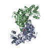
| Deposited unit | 
| |||||||||
|---|---|---|---|---|---|---|---|---|---|---|
| 1 | 
| |||||||||
| Unit cell |
| |||||||||
| Components on special symmetry positions |
| |||||||||
| Details | The biological assembly is a dimer |
-Components
| #1: Protein | Mass: 49091.797 Da / Num. of mol.: 1 / Fragment: RESIDUES 1-435 Source method: isolated from a genetically manipulated source Source: (gene. exp.)  References: UniProt: P37821, 1-aminocyclopropane-1-carboxylate synthase |
|---|---|
| #2: Chemical | ChemComp-PPG / (  Mass: 389.298 Da / Num. of mol.: 1 / Source method: obtained synthetically / Formula: C14H20N3O8P Mass: 389.298 Da / Num. of mol.: 1 / Source method: obtained synthetically / Formula: C14H20N3O8P |
| #3: Chemical | ChemComp-MRD / (  Mass: 118.174 Da / Num. of mol.: 1 / Source method: obtained synthetically / Formula: C6H14O2 / Comment: precipitant*YM Mass: 118.174 Da / Num. of mol.: 1 / Source method: obtained synthetically / Formula: C6H14O2 / Comment: precipitant*YM |
| #4: Water | ChemComp-HOH /  Mass: 18.015 Da / Num. of mol.: 332 / Source method: isolated from a natural source / Formula: H2O Mass: 18.015 Da / Num. of mol.: 332 / Source method: isolated from a natural source / Formula: H2O |
-Experimental details
-Experiment
| Experiment | Method: X-RAY DIFFRACTION / Number of used crystals: 1 |
|---|
- Sample preparation
| Crystal | Density Matthews: 2.07 Å3/Da / Density % sol: 40.63 % | ||||||||||||||||||||||||||||||||||||||||||||||||
|---|---|---|---|---|---|---|---|---|---|---|---|---|---|---|---|---|---|---|---|---|---|---|---|---|---|---|---|---|---|---|---|---|---|---|---|---|---|---|---|---|---|---|---|---|---|---|---|---|---|
| Crystal grow | Temperature: 293 K / Method: vapor diffusion, sitting drop / pH: 6.5 Details: MPD, MES, pH 6.5, VAPOR DIFFUSION, SITTING DROP, temperature 293K | ||||||||||||||||||||||||||||||||||||||||||||||||
| Crystal grow | *PLUS pH: 7.9 | ||||||||||||||||||||||||||||||||||||||||||||||||
| Components of the solutions | *PLUS
|
-Data collection
| Diffraction | Mean temperature: 100 K |
|---|---|
| Diffraction source | Source: SYNCHROTRON / Site: ESRF  / Beamline: BM1A / Wavelength: 0.79955 Å / Beamline: BM1A / Wavelength: 0.79955 Å |
| Detector | Type: MARRESEARCH / Detector: IMAGE PLATE / Date: Jun 18, 2000 |
| Radiation | Monochromator: SAGITALLY FOCUSED Si(111) / Protocol: SINGLE WAVELENGTH / Monochromatic (M) / Laue (L): M / Scattering type: x-ray |
| Radiation wavelength | Wavelength: 0.79955 Å / Relative weight: 1 |
| Reflection | Resolution: 1.6→15 Å / Num. obs: 50871 / % possible obs: 95.2 % / Observed criterion σ(I): -3 / Redundancy: 2.8 % / Biso Wilson estimate: 18.7 Å2 / Rmerge(I) obs: 0.052 / Net I/σ(I): 16.5 |
| Reflection shell | Resolution: 1.6→1.64 Å / Rmerge(I) obs: 0.281 / Mean I/σ(I) obs: 2.9 / Num. unique all: 2684 / % possible all: 76 |
| Reflection | *PLUS Highest resolution: 1.6 Å / Lowest resolution: 15 Å / Num. obs: 50540 |
| Reflection shell | *PLUS % possible obs: 76 % |
- Processing
| Software |
| ||||||||||||||||||||
|---|---|---|---|---|---|---|---|---|---|---|---|---|---|---|---|---|---|---|---|---|---|
| Refinement | Method to determine structure: MOLECULAR REPLACEMENT Starting model: PDB ENTRY 1B8G Resolution: 1.6→14.924 Å / Isotropic thermal model: ISOTROPIC / Cross valid method: THROUGHOUT / σ(F): 0 / Stereochemistry target values: Engh & Huber / Details: Used bulk solvent correction (CNS 1.0)
| ||||||||||||||||||||
| Displacement parameters | Biso mean: 23.5 Å2
| ||||||||||||||||||||
| Refine analyze |
| ||||||||||||||||||||
| Refinement step | Cycle: LAST / Resolution: 1.6→14.924 Å
| ||||||||||||||||||||
| Refine LS restraints |
| ||||||||||||||||||||
| LS refinement shell | Resolution: 1.6→1.66 Å
| ||||||||||||||||||||
| Refinement | *PLUS Highest resolution: 1.6 Å / Lowest resolution: 15 Å / % reflection Rfree: 5 % | ||||||||||||||||||||
| Solvent computation | *PLUS | ||||||||||||||||||||
| Displacement parameters | *PLUS | ||||||||||||||||||||
| Refine LS restraints | *PLUS
|
