 Movie
Movie Controller

[English] 日本語
 Yorodumi
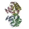
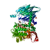
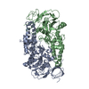
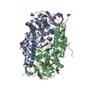
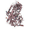
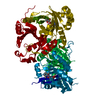
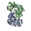
- PDB-1b4u: PROTOCATECHUATE 4,5-DIOXYGENASE (LIGAB) IN COMPLEX WITH PROTOCATE... -
+ Open data

- Basic information
| Entry | Database: PDB / ID: 1b4u | ||||||
|---|---|---|---|---|---|---|---|
| Title | PROTOCATECHUATE 4,5-DIOXYGENASE (LIGAB) IN COMPLEX WITH PROTOCATECHUATE (PCA) | ||||||
 Components Components | (PROTOCATECHUATE 4,5-DIOXYGENASE) x 2 | ||||||
 Keywords Keywords | DIOXYGENASE / EXTRADIOL TYPE DIOXYGENASE / PROTOCATECHUATE / NON-HEME IRON PROTEIN | ||||||
| Function / homology |  Function and homology information Function and homology informationprotocatechuate 4,5-dioxygenase / protocatechuate 4,5-dioxygenase activity / ferrous iron binding Similarity search - Function | ||||||
| Biological species |  Sphingomonas paucimobilis (bacteria) Sphingomonas paucimobilis (bacteria) | ||||||
| Method |  X-RAY DIFFRACTION / MIRAS / Resolution: 2.2 Å X-RAY DIFFRACTION / MIRAS / Resolution: 2.2 Å | ||||||
 Authors Authors | Sugimoto, K. / Senda, T. / Mitsui, Y. | ||||||
 Citation Citation | Journal: Structure Fold.Des. / Year: 1999 Title: Crystal structure of an aromatic ring opening dioxygenase LigAB, a protocatechuate 4,5-dioxygenase, under aerobic conditions. Authors: Sugimoto, K. / Senda, T. / Aoshima, H. / Masai, E. / Fukuda, M. / Mitsui, Y. | ||||||
| History |
|
- Structure visualization
| Structure viewer | Molecule: MolmilJmol/JSmol |
|---|
- Downloads & links
-Download
| PDBx/mmCIF format | 1b4u.cif.gz | 177.1 KB | Display | PDBx/mmCIF format |
|---|---|---|---|---|
| PDB format | pdb1b4u.ent.gz | 141.3 KB | Display | PDB format |
| PDBx/mmJSON format | 1b4u.json.gz | Tree view | PDBx/mmJSON format | |
| Others |  Other downloads Other downloads |
-Validation report
| Arichive directory | https://data.pdbj.org/pub/pdb/validation_reports/b4/1b4uftp://data.pdbj.org/pub/pdb/validation_reports/b4/1b4u | HTTPS FTP |
|---|
-Related structure data


-Links
 PDBj
PDBj- Assembly
| Deposited unit | 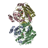
| ||||||||||||
|---|---|---|---|---|---|---|---|---|---|---|---|---|---|
| 1 |
| ||||||||||||
| Unit cell |
| ||||||||||||
| Noncrystallographic symmetry (NCS) | NCS oper:
|
-Components
| #1: Protein | Mass: 15567.516 Da / Num. of mol.: 2 / Fragment: CHAIN A, C, ALPHA CHAIN, CHAIN B, D, BETA CHAIN Source method: isolated from a genetically manipulated source Source: (gene. exp.) Sphingomonas paucimobilis (bacteria) / Strain: SYK-6 / Production host: References: UniProt: P22635, protocatechuate 4,5-dioxygenase #2: Protein | Mass: 33322.996 Da / Num. of mol.: 2 / Fragment: CHAIN A, C, ALPHA CHAIN, CHAIN B, D, BETA CHAIN Source method: isolated from a genetically manipulated source Source: (gene. exp.) Sphingomonas paucimobilis (bacteria) / Strain: SYK-6 / Production host: References: UniProt: P22636, protocatechuate 4,5-dioxygenase #3: Chemical |   Mass: 55.845 Da / Num. of mol.: 2 / Source method: obtained synthetically / Formula: Fe Mass: 55.845 Da / Num. of mol.: 2 / Source method: obtained synthetically / Formula: Fe#4: Chemical | 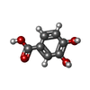  Mass: 154.120 Da / Num. of mol.: 2 / Source method: obtained synthetically / Formula: C7H6O4 Mass: 154.120 Da / Num. of mol.: 2 / Source method: obtained synthetically / Formula: C7H6O4#5: Water | ChemComp-HOH / |  Mass: 18.015 Da / Num. of mol.: 189 / Source method: isolated from a natural source / Formula: H2O Mass: 18.015 Da / Num. of mol.: 189 / Source method: isolated from a natural source / Formula: H2O |
|---|
-Experimental details
-Experiment
| Experiment | Method: X-RAY DIFFRACTION / Number of used crystals: 1 |
|---|
- Sample preparation
| Crystal | Density Matthews: 2.66 Å3/Da / Density % sol: 53.76 % | |||||||||||||||||||||||||
|---|---|---|---|---|---|---|---|---|---|---|---|---|---|---|---|---|---|---|---|---|---|---|---|---|---|---|
| Crystal grow | pH: 7.4 / Details: pH 7.4 | |||||||||||||||||||||||||
| Crystal | *PLUS Density % sol: 53.4 % | |||||||||||||||||||||||||
| Crystal grow | *PLUS Temperature: 20 ℃ / pH: 7.5 / Method: vapor diffusion, hanging drop / Details: Sugimoto, K., (1999) Protein Peptide Lett., 6, 55. | |||||||||||||||||||||||||
| Components of the solutions | *PLUS
|
-Data collection
| Diffraction | Mean temperature: 300 K |
|---|---|
| Diffraction source | Source: ROTATING ANODE / Type: RIGAKU RUH2R / Wavelength: 1.5418 |
| Detector | Type: RIGAKU / Detector: IMAGE PLATE / Date: Jul 29, 1998 / Details: MIRRORS |
| Radiation | Monochromatic (M) / Laue (L): M / Scattering type: x-ray |
| Radiation wavelength | Wavelength: 1.5418 Å / Relative weight: 1 |
| Reflection | Resolution: 2.2→60 Å / Num. obs: 45587 / % possible obs: 91 % / Observed criterion σ(I): 1 / Redundancy: 2 % / Rmerge(I) obs: 0.055 / Net I/σ(I): 11 |
| Reflection | *PLUS Num. measured all: 91377 |
- Processing
| Software | Name: REFMAC / Classification: refinement | ||||||||||||||||||||||||||||||||||||||||||||||||||||||||||||||||||||||||||||||||||||
|---|---|---|---|---|---|---|---|---|---|---|---|---|---|---|---|---|---|---|---|---|---|---|---|---|---|---|---|---|---|---|---|---|---|---|---|---|---|---|---|---|---|---|---|---|---|---|---|---|---|---|---|---|---|---|---|---|---|---|---|---|---|---|---|---|---|---|---|---|---|---|---|---|---|---|---|---|---|---|---|---|---|---|---|---|---|
| Refinement | Method to determine structure: MIRAS / Resolution: 2.2→60 Å / Cross valid method: THROUGHOUT / σ(F): 1
| ||||||||||||||||||||||||||||||||||||||||||||||||||||||||||||||||||||||||||||||||||||
| Refinement step | Cycle: LAST / Resolution: 2.2→60 Å
| ||||||||||||||||||||||||||||||||||||||||||||||||||||||||||||||||||||||||||||||||||||
| Refine LS restraints |
| ||||||||||||||||||||||||||||||||||||||||||||||||||||||||||||||||||||||||||||||||||||
| Refinement | *PLUS Rfactor obs: 0.161 / Rfactor Rfree: 0.22 | ||||||||||||||||||||||||||||||||||||||||||||||||||||||||||||||||||||||||||||||||||||
| Solvent computation | *PLUS | ||||||||||||||||||||||||||||||||||||||||||||||||||||||||||||||||||||||||||||||||||||
| Displacement parameters | *PLUS |
