| 登録情報 | データベース: PDB / ID: 1m61
|
|---|
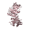
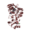
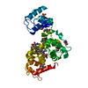
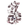
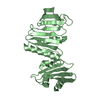
| タイトル | Crystal structure of the apo SH2 domains of ZAP-70 |
|---|
 要素 要素 | TYROSINE-PROTEIN KINASE ZAP-70 |
|---|
 キーワード キーワード | TRANSFERASE / apo form |
|---|
| 機能・相同性 |  機能・相同性情報 機能・相同性情報
T cell aggregation / positive regulation of alpha-beta T cell proliferation / negative thymic T cell selection / beta selection / positive thymic T cell selection / positive regulation of alpha-beta T cell differentiation / positive regulation of T cell differentiation / T cell receptor complex / Translocation of ZAP-70 to Immunological synapse / B cell activation ...T cell aggregation / positive regulation of alpha-beta T cell proliferation / negative thymic T cell selection / beta selection / positive thymic T cell selection / positive regulation of alpha-beta T cell differentiation / positive regulation of T cell differentiation / T cell receptor complex / Translocation of ZAP-70 to Immunological synapse / B cell activation / Generation of second messenger molecules / RHOH GTPase cycle / immunological synapse / T cell differentiation / Nuclear events stimulated by ALK signaling in cancer / T cell migration / phosphotyrosine residue binding / positive regulation of calcium-mediated signaling / T cell activation / peptidyl-tyrosine phosphorylation / non-membrane spanning protein tyrosine kinase activity / non-specific protein-tyrosine kinase / calcium-mediated signaling / cell-cell junction / T cell receptor signaling pathway / protein tyrosine kinase activity / adaptive immune response / protein phosphorylation / intracellular signal transduction / immune response / ATP binding / plasma membrane / cytoplasm / cytosol類似検索 - 分子機能 Syk Kinase; Chain A, domain 2 / Syk Kinase; Chain A, domain 2 / Tyrosine-protein kinase, non-receptor SYK/ZAP-70 / Tyrosine-protein kinase SYK/ZAP-70, inter-SH2 domain superfamily / SYK/ZAP-70, N-terminal SH2 domain / SH2 domain / SHC Adaptor Protein / : / SH2 domain / Src homology 2 (SH2) domain profile. ...Syk Kinase; Chain A, domain 2 / Syk Kinase; Chain A, domain 2 / Tyrosine-protein kinase, non-receptor SYK/ZAP-70 / Tyrosine-protein kinase SYK/ZAP-70, inter-SH2 domain superfamily / SYK/ZAP-70, N-terminal SH2 domain / SH2 domain / SHC Adaptor Protein / : / SH2 domain / Src homology 2 (SH2) domain profile. / Src homology 2 domains / SH2 domain / SH2 domain superfamily / Tyrosine-protein kinase, catalytic domain / Tyrosine kinase, catalytic domain / Tyrosine protein kinases specific active-site signature. / Tyrosine-protein kinase, active site / Serine-threonine/tyrosine-protein kinase, catalytic domain / Protein tyrosine and serine/threonine kinase / Protein kinase, ATP binding site / Protein kinases ATP-binding region signature. / Protein kinase domain profile. / Protein kinase domain / Protein kinase-like domain superfamily / 2-Layer Sandwich / Orthogonal Bundle / Mainly Alpha / Alpha Beta類似検索 - ドメイン・相同性 PHOSPHATE ION / Tyrosine-protein kinase ZAP-70類似検索 - 構成要素 |
|---|
| 生物種 |  Homo sapiens (ヒト) Homo sapiens (ヒト) |
|---|
| 手法 |  X線回折 / シンクロトロン / 分子置換 / 解像度: 2.5 Å X線回折 / シンクロトロン / 分子置換 / 解像度: 2.5 Å |
|---|
 データ登録者 データ登録者 | Folmer, R.H.A. / Geschwindner, S. / Xue, Y. |
|---|
 引用 引用 | ジャーナル: Biochemistry / 年: 2002
タイトル: Crystal structure and NMR studies of the apo SH2 domains of ZAP-70: two bikes rather than a tandem
著者: Folmer, R.H.A. / Geschwindner, S. / Xue, Y. |
|---|
| 履歴 | | 登録 | 2002年7月11日 | 登録サイト: RCSB / 処理サイト: RCSB |
|---|
| 改定 1.0 | 2003年7月15日 | Provider: repository / タイプ: Initial release |
|---|
| 改定 1.1 | 2008年4月28日 | Group: Version format compliance |
|---|
| 改定 1.2 | 2011年7月13日 | Group: Version format compliance |
|---|
| 改定 1.3 | 2018年3月7日 | Group: Data collection / カテゴリ: diffrn_source / Item: _diffrn_source.pdbx_synchrotron_site |
|---|
| 改定 1.4 | 2024年2月14日 | Group: Data collection / Database references ...Data collection / Database references / Derived calculations / Refinement description
カテゴリ: chem_comp_atom / chem_comp_bond ...chem_comp_atom / chem_comp_bond / database_2 / pdbx_initial_refinement_model / struct_ref_seq_dif / struct_site
Item: _database_2.pdbx_DOI / _database_2.pdbx_database_accession ..._database_2.pdbx_DOI / _database_2.pdbx_database_accession / _struct_ref_seq_dif.details / _struct_site.pdbx_auth_asym_id / _struct_site.pdbx_auth_comp_id / _struct_site.pdbx_auth_seq_id |
|---|
|
|---|
 ムービー
ムービー コントローラー


 データを開く
 基本情報
基本情報 構造の表示
構造の表示 ダウンロードとリンク
 その他のダウンロード
 PDBj
PDBj


 集合体
集合体


 分子量: 94.971 Da / 分子数: 1 / 由来タイプ: 合成 / 式: PO4
 分子量: 18.015 Da / 分子数: 36 / 由来タイプ: 天然 / 式: H2O
 試料調製
試料調製 / ビームライン: I711 / 波長: 1.0158 Å
 解析
解析