 Movie
Movie Controller


+ Open data

- Basic information
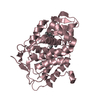
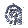
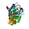
| Entry | Database: PDB / ID: 1m4r | ||||||
|---|---|---|---|---|---|---|---|
| Title | CRYSTAL STRUCTURE OF RECOMBINANT HUMAN INTERLEUKIN-22 | ||||||
 Components Components | Interleukin-22 | ||||||
 Keywords Keywords | CYTOKINE / interleukin-22 (IL-22) / interleukin-10 (IL-10) / interferon-gamma / cytokines | ||||||
| Function / homology |  Function and homology information Function and homology informationinterleukin-22 receptor binding / Interleukin-20 family signaling / response to glucocorticoid / cytokine activity / acute-phase response / negative regulation of inflammatory response / Signaling by ALK fusions and activated point mutants / inflammatory response / : / extracellular region Similarity search - Function | ||||||
| Biological species |  Homo sapiens (human) Homo sapiens (human) | ||||||
| Method |  X-RAY DIFFRACTION / SYNCHROTRON / SIRAS / Resolution: 2 Å X-RAY DIFFRACTION / SYNCHROTRON / SIRAS / Resolution: 2 Å | ||||||
 Authors Authors | Nagem, R.A.P. / Colau, D. / Dumoutier, L. / Renauld, J.-C. / Ogata, C. / Polikarpov, I. | ||||||
 Citation Citation | Journal: Structure / Year: 2002 Title: Crystal Structure of Recombinant Human Interleukin-22 Authors: Nagem, R.A.P. / Colau, D. / Dumoutier, L. / Renauld, J.-C. / Ogata, C. / Polikarpov, I. #1: Journal: Acta Crystallogr.,Sect.D / Year: 2002Title: Crystallization and Synchrotron X-ray Diffraction Studies of Human Interleukin-22 Authors: Nagem, R.A.P. / Lucchesi, K.W. / Colau, D. / Dumoutier, L. / Renauld, J.-C. / Polikarpov, I. | ||||||
| History |
|
- Structure visualization
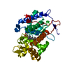
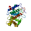
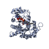
| Structure viewer | Molecule: MolmilJmol/JSmol |
|---|
- Downloads & links
-Download
| PDBx/mmCIF format | 1m4r.cif.gz | 72.1 KB | Display | PDBx/mmCIF format |
|---|---|---|---|---|
| PDB format | pdb1m4r.ent.gz | 54.7 KB | Display | PDB format |
| PDBx/mmJSON format | 1m4r.json.gz | Tree view | PDBx/mmJSON format | |
| Others |  Other downloads Other downloads |
-Validation report
| Arichive directory | https://data.pdbj.org/pub/pdb/validation_reports/m4/1m4rftp://data.pdbj.org/pub/pdb/validation_reports/m4/1m4r | HTTPS FTP |
|---|
-Related structure data
| Similar structure data |
|---|


-Links
 PDBj
PDBj


- Assembly
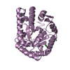
| Deposited unit | 
| ||||||||
|---|---|---|---|---|---|---|---|---|---|
| 1 |
| ||||||||
| Unit cell |
|
-Components
| #1: Protein | Mass: 17155.730 Da / Num. of mol.: 2 Source method: isolated from a genetically manipulated source Source: (gene. exp.) Homo sapiens (human) / Plasmid: pET3A / Production host:  #2: Water | ChemComp-HOH / |  Mass: 18.015 Da / Num. of mol.: 189 / Source method: isolated from a natural source / Formula: H2O Mass: 18.015 Da / Num. of mol.: 189 / Source method: isolated from a natural source / Formula: H2OHas protein modification | Y | |
|---|
-Experimental details
-Experiment
| Experiment | Method: X-RAY DIFFRACTION / Number of used crystals: 1 |
|---|
- Sample preparation
| Crystal | Density Matthews: 2.02 Å3/Da / Density % sol: 39 % | ||||||||||||||||||||||||||||||
|---|---|---|---|---|---|---|---|---|---|---|---|---|---|---|---|---|---|---|---|---|---|---|---|---|---|---|---|---|---|---|---|
| Crystal grow | Temperature: 300 K / Method: vapor diffusion, hanging drop / pH: 7.5 Details: 0.9 sodium tartrate, TRITON X-100 detergent, 0.1 M HEPES, pH 7.5, VAPOR DIFFUSION, HANGING DROP, temperature 300K | ||||||||||||||||||||||||||||||
| Crystal grow | *PLUS pH: 5.4 / Method: vapor diffusion, hanging drop | ||||||||||||||||||||||||||||||
| Components of the solutions | *PLUS
|
-Data collection
| Diffraction | Mean temperature: 100 K |
|---|---|
| Diffraction source | Source: SYNCHROTRON / Site: LNLS  / Beamline: D03B-MX1 / Wavelength: 1.54 Å / Beamline: D03B-MX1 / Wavelength: 1.54 Å |
| Detector | Type: MARRESEARCH / Detector: IMAGE PLATE / Date: Jan 1, 2001 |
| Radiation | Monochromator: Si (111) single crystal / Protocol: SINGLE WAVELENGTH / Monochromatic (M) / Laue (L): M / Scattering type: x-ray |
| Radiation wavelength | Wavelength: 1.54 Å / Relative weight: 1 |
| Reflection | Resolution: 2→21.72 Å / Num. all: 18125 / Num. obs: 16349 / % possible obs: 92 % / Observed criterion σ(F): 0 / Observed criterion σ(I): 0 / Biso Wilson estimate: 16.1 Å2 / Rmerge(I) obs: 0.082 / Net I/σ(I): 14.5 |
| Reflection shell | Resolution: 2→2.05 Å / Rmerge(I) obs: 0.35 / Mean I/σ(I) obs: 3.8 / % possible all: 82.3 |
| Reflection | *PLUS Lowest resolution: 21.7 Å / Num. obs: 16382 / % possible obs: 92.7 % / Redundancy: 3.8 % / Num. measured all: 61846 |
| Reflection shell | *PLUS % possible obs: 82.3 % / Redundancy: 3.4 % / Rmerge(I) obs: 0.35 |
- Processing
| Software |
| ||||||||||||||||||||||||||||||||||||
|---|---|---|---|---|---|---|---|---|---|---|---|---|---|---|---|---|---|---|---|---|---|---|---|---|---|---|---|---|---|---|---|---|---|---|---|---|---|
| Refinement | Method to determine structure: SIRAS / Resolution: 2→21.72 Å / Rfactor Rfree error: 0.008 / Data cutoff high absF: 1118732.49 / Data cutoff high rms absF: 1118732.49 / Data cutoff low absF: 0 / Isotropic thermal model: RESTRAINED / Cross valid method: THROUGHOUT / σ(F): 0 / Stereochemistry target values: Engh & Huber
| ||||||||||||||||||||||||||||||||||||
| Solvent computation | Solvent model: FLAT MODEL / Bsol: 56.9772 Å2 / ksol: 0.38446 e/Å3 | ||||||||||||||||||||||||||||||||||||
| Displacement parameters | Biso mean: 25.1 Å2
| ||||||||||||||||||||||||||||||||||||
| Refine analyze |
| ||||||||||||||||||||||||||||||||||||
| Refinement step | Cycle: LAST / Resolution: 2→21.72 Å
| ||||||||||||||||||||||||||||||||||||
| Refine LS restraints |
| ||||||||||||||||||||||||||||||||||||
| LS refinement shell | Resolution: 2→2.07 Å / Rfactor Rfree error: 0 / Total num. of bins used: 6
| ||||||||||||||||||||||||||||||||||||
| Xplor file |
| ||||||||||||||||||||||||||||||||||||
| Refinement | *PLUS Highest resolution: 2 Å / Lowest resolution: 21.7 Å / Num. reflection all: 15684 / Num. reflection obs: 14892 / Num. reflection Rfree: 792 / Rfactor Rfree: 0.22 | ||||||||||||||||||||||||||||||||||||
| Solvent computation | *PLUS | ||||||||||||||||||||||||||||||||||||
| Displacement parameters | *PLUS | ||||||||||||||||||||||||||||||||||||
| Refine LS restraints | *PLUS
|
