 Movie
Movie Controller

[English] 日本語
 Yorodumi
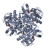
- PDB-1m41: Crystal structure of Escherichia coli alkanesulfonate monooxygena... -
+ Open data

- Basic information
| Entry | Database: PDB / ID: 1m41 | ||||||
|---|---|---|---|---|---|---|---|
| Title | Crystal structure of Escherichia coli alkanesulfonate monooxygenase SsuD at 2.3 A resolution | ||||||
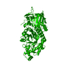
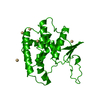
 Components Components | FMNH2-dependent alkanesulfonate monooxygenase | ||||||
 Keywords Keywords | OXIDOREDUCTASE / FMNH2-dependent monooxygenase / SsuD / TIM-barrel / sulfate starvation / sulfur assimilation / desulfonation / alkanesulfonate / oxygenase / monooxygenase | ||||||
| Function / homology |  Function and homology information Function and homology informationalkanesulfonate monooxygenase complex / alkanesulfonate monooxygenase / cellular response to sulfur starvation / alkanesulfonate monooxygenase activity / alkanesulfonate catabolic process / response to heat / protein homotetramerization / identical protein binding Similarity search - Function | ||||||
| Biological species |  | ||||||
| Method |  X-RAY DIFFRACTION / SYNCHROTRON / MAD / Resolution: 2.3 Å X-RAY DIFFRACTION / SYNCHROTRON / MAD / Resolution: 2.3 Å | ||||||
 Authors Authors | Eichhorn, E. / Davey, C.A. / Sargent, D.F. / Leisinger, T. / Richmond, T.J. | ||||||
 Citation Citation | Journal: J.mol.biol. / Year: 2002 Title: Crystal Structure of Escherichia coli Alkanesulfonate Monooxygenase SsuD Authors: Eichhorn, E. / Davey, C.A. / Sargent, D.F. / Leisinger, T. / Richmond, T.J. | ||||||
| History |
|
- Structure visualization
| Structure viewer | Molecule: MolmilJmol/JSmol |
|---|
- Downloads & links
-Download
| PDBx/mmCIF format | 1m41.cif.gz | 144.4 KB | Display | PDBx/mmCIF format |
|---|---|---|---|---|
| PDB format | pdb1m41.ent.gz | 113.5 KB | Display | PDB format |
| PDBx/mmJSON format | 1m41.json.gz | Tree view | PDBx/mmJSON format | |
| Others |  Other downloads Other downloads |
-Validation report
| Summary document | 1m41_validation.pdf.gz | 376.4 KB | Display | wwPDB validaton report |
|---|---|---|---|---|
| Full document | 1m41_full_validation.pdf.gz | 403.8 KB | Display | |
| Data in XML | 1m41_validation.xml.gz | 17.3 KB | Display | |
| Data in CIF | 1m41_validation.cif.gz | 27.3 KB | Display | |
| Arichive directory | https://data.pdbj.org/pub/pdb/validation_reports/m4/1m41ftp://data.pdbj.org/pub/pdb/validation_reports/m4/1m41 | HTTPS FTP |
-Related structure data
| Similar structure data |
|---|

-Links
 PDBj
PDBj
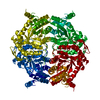
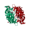
- Assembly
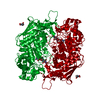
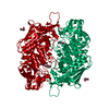
| Deposited unit | 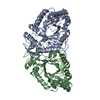
| ||||||||
|---|---|---|---|---|---|---|---|---|---|
| 1 | 
| ||||||||
| Unit cell |
| ||||||||
| Components on special symmetry positions |
| ||||||||
| Details | The second part of the biological assembly is generated by the two fold axis: -x, y, -z+1/2 |
-Components
| #1: Protein | Mass: 41651.906 Da / Num. of mol.: 2 Source method: isolated from a genetically manipulated source Source: (gene. exp.) References: UniProt: P80645, Oxidoreductases; Acting on the CH-OH group of donors #2: Water | ChemComp-HOH / |  Mass: 18.015 Da / Num. of mol.: 310 / Source method: isolated from a natural source / Formula: H2O Mass: 18.015 Da / Num. of mol.: 310 / Source method: isolated from a natural source / Formula: H2O |
|---|
-Experimental details
-Experiment
| Experiment | Method: X-RAY DIFFRACTION / Number of used crystals: 1 |
|---|
- Sample preparation
| Crystal | Density Matthews: 3 Å3/Da / Density % sol: 60 % | ||||||||||||||||||||
|---|---|---|---|---|---|---|---|---|---|---|---|---|---|---|---|---|---|---|---|---|---|
| Crystal grow | Temperature: 297 K / Method: vapor diffusion, sitting drop / pH: 7 Details: sodium formate, pH 7, VAPOR DIFFUSION, SITTING DROP, temperature 297K | ||||||||||||||||||||
| Crystal grow | *PLUS | ||||||||||||||||||||
| Components of the solutions | *PLUS
|
-Data collection
| Diffraction |
| ||||||||||||||||||
|---|---|---|---|---|---|---|---|---|---|---|---|---|---|---|---|---|---|---|---|
| Diffraction source |
| ||||||||||||||||||
| Detector |
| ||||||||||||||||||
| Radiation |
| ||||||||||||||||||
| Radiation wavelength |
| ||||||||||||||||||
| Reflection | Resolution: 2.3→25 Å / Num. all: 44869 / Num. obs: 44511 / % possible obs: 99.2 % / Observed criterion σ(F): 1 / Observed criterion σ(I): 1 / Redundancy: 4.7 % / Biso Wilson estimate: 42.4 Å2 / Rmerge(I) obs: 0.062 / Net I/σ(I): 7.8 | ||||||||||||||||||
| Reflection shell | Resolution: 2.3→2.41 Å / Redundancy: 3.5 % / Rmerge(I) obs: 0.432 / Mean I/σ(I) obs: 1.6 / Num. unique all: 6348 / % possible all: 95.5 | ||||||||||||||||||
| Reflection | *PLUS Lowest resolution: 24.7 Å / Num. obs: 44869 / % possible obs: 97.3 % / Num. measured all: 752595 | ||||||||||||||||||
| Reflection shell | *PLUS % possible obs: 95.5 % |

- Processing
| Software |
| |||||||||||||||||||||||||
|---|---|---|---|---|---|---|---|---|---|---|---|---|---|---|---|---|---|---|---|---|---|---|---|---|---|---|
| Refinement | Method to determine structure: MAD / Resolution: 2.3→25 Å / Rfactor Rfree error: 0.006 / Isotropic thermal model: restrained / Cross valid method: THROUGHOUT / σ(F): 0 / Stereochemistry target values: Engh & Huber
| |||||||||||||||||||||||||
| Displacement parameters | Biso mean: 55.8 Å2
| |||||||||||||||||||||||||
| Refine analyze |
| |||||||||||||||||||||||||
| Refinement step | Cycle: LAST / Resolution: 2.3→25 Å
| |||||||||||||||||||||||||
| Refine LS restraints |
| |||||||||||||||||||||||||
| LS refinement shell | Resolution: 2.3→2.38 Å / Rfactor Rfree error: 0.03 / Total num. of bins used: 10
| |||||||||||||||||||||||||
| Refinement | *PLUS Lowest resolution: 25 Å / % reflection Rfree: 2.5 % | |||||||||||||||||||||||||
| Solvent computation | *PLUS | |||||||||||||||||||||||||
| Displacement parameters | *PLUS | |||||||||||||||||||||||||
| Refine LS restraints | *PLUS
|
