[English] 日本語
 Yorodumi
- PDB-1m3c: Solution structure of a circular form of the N-terminal SH3 domai... -
+ Open data
- Basic information
| Entry | Database: PDB / ID: 1m3c | ||||||
|---|---|---|---|---|---|---|---|
| Title | Solution structure of a circular form of the N-terminal SH3 domain (E132C, E133G, R191G mutant) from oncogene protein c-Crk | ||||||
 Components Components | Proto-oncogene C-crk | ||||||
 Keywords Keywords | PROTEIN BINDING / SH3 / SH3 DOMAIN / CIRCULAR PROTEIN / CYCLIZED PROTEIN / ADAPTOR PROTEIN | ||||||
| Function / homology |  Function and homology information Function and homology informationPTK6 Regulates RHO GTPases, RAS GTPase and MAP kinases / ARMS-mediated activation / MET activates RAP1 and RAC1 / response to hepatocyte growth factor / MET receptor recycling / helper T cell diapedesis / cerebellar neuron development / response to cholecystokinin / Downstream signal transduction / cellular response to endothelin ...PTK6 Regulates RHO GTPases, RAS GTPase and MAP kinases / ARMS-mediated activation / MET activates RAP1 and RAC1 / response to hepatocyte growth factor / MET receptor recycling / helper T cell diapedesis / cerebellar neuron development / response to cholecystokinin / Downstream signal transduction / cellular response to endothelin / regulation of leukocyte migration / postsynaptic specialization assembly / protein phosphorylated amino acid binding / regulation of T cell migration / p130Cas linkage to MAPK signaling for integrins / response to peptide / regulation of dendrite development / Regulation of signaling by CBL / negative regulation of wound healing / Regulation of actin dynamics for phagocytic cup formation / response to yeast / positive regulation of skeletal muscle acetylcholine-gated channel clustering / reelin-mediated signaling pathway / VEGFA-VEGFR2 Pathway / negative regulation of cell motility / negative regulation of natural killer cell mediated cytotoxicity / protein localization to membrane / regulation of GTPase activity / positive regulation of smooth muscle cell migration / regulation of cell adhesion mediated by integrin / enzyme-linked receptor protein signaling pathway / cellular response to insulin-like growth factor stimulus / establishment of cell polarity / dendrite development / positive regulation of Rac protein signal transduction / ephrin receptor signaling pathway / cellular response to transforming growth factor beta stimulus / ephrin receptor binding / cytoskeletal protein binding / insulin-like growth factor receptor binding / phosphotyrosine residue binding / positive regulation of substrate adhesion-dependent cell spreading / cellular response to nitric oxide / signaling adaptor activity / SH2 domain binding / protein tyrosine kinase binding / cell chemotaxis / regulation of actin cytoskeleton organization / hippocampus development / neuromuscular junction / lipid metabolic process / response to hydrogen peroxide / cellular response to nerve growth factor stimulus / cerebral cortex development / positive regulation of JNK cascade / SH3 domain binding / neuron migration / regulation of cell shape / actin cytoskeleton / signaling receptor complex adaptor activity / actin cytoskeleton organization / scaffold protein binding / cell population proliferation / ubiquitin protein ligase binding / protein-containing complex / membrane / plasma membrane / cytoplasm Similarity search - Function | ||||||
| Biological species |  | ||||||
| Method | SOLUTION NMR / Simulated annealing, torsion angle dynamics | ||||||
 Authors Authors | Schumann, F.H. / Varadan, R. / Tayakuniyil, P.P. / Hall, J.B. / Camarero, J.A. / Fushman, D. | ||||||
 Citation Citation |  Journal: To be Published Journal: To be PublishedTitle: Changing protein backbone topology: Structural and dynamic consequences of the backbone cyclization in SH3 domain Authors: Schumann, F.H. / Varadan, R. / Tayakuniyil, P.P. / Hall, J.B. / Camarero, J.A. / Fushman, D. #1:  Journal: J.Mol.Biol. / Year: 2001 Journal: J.Mol.Biol. / Year: 2001Title: Rescuing a destabilized protein fold through backbone cyclization Authors: Camarero, J.A. / Fushman, D. / Sato, S. / Giriat, I. / Cowburn, D. / Raleigh, D.P. / Muir, T.W. | ||||||
| History |
|
- Structure visualization
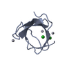
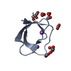
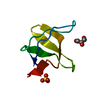
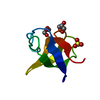
| Structure viewer | Molecule:  Molmil Molmil Jmol/JSmol Jmol/JSmol |
|---|
- Downloads & links
- Download
| PDBx/mmCIF format |  1m3c.cif.gz 1m3c.cif.gz | 377.9 KB | Display |  PDBx/mmCIF format PDBx/mmCIF format |
|---|---|---|---|---|
| PDB format |  pdb1m3c.ent.gz pdb1m3c.ent.gz | 314.7 KB | Display |  PDB format PDB format |
| PDBx/mmJSON format |  1m3c.json.gz 1m3c.json.gz | Tree view |  PDBx/mmJSON format PDBx/mmJSON format | |
| Others |  Other downloads Other downloads |
-Validation report
| Arichive directory |  https://data.pdbj.org/pub/pdb/validation_reports/m3/1m3c https://data.pdbj.org/pub/pdb/validation_reports/m3/1m3c ftp://data.pdbj.org/pub/pdb/validation_reports/m3/1m3c ftp://data.pdbj.org/pub/pdb/validation_reports/m3/1m3c | HTTPS FTP |
|---|
-Related structure data
- Links
Links
- Assembly
| Deposited unit | 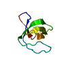
| |||||||||
|---|---|---|---|---|---|---|---|---|---|---|
| 1 |
| |||||||||
| NMR ensembles |
|
- Components
| #1: Protein | Mass: 7031.783 Da / Num. of mol.: 1 / Fragment: N-TERMINAL SH3 DOMAIN (residues 132-191) / Mutation: R191G, added G133, C132 Source method: isolated from a genetically manipulated source Source: (gene. exp.)   |
|---|---|
| Has protein modification | Y |
-Experimental details
-Experiment
| Experiment | Method: SOLUTION NMR | ||||||||||||||||||||||||
|---|---|---|---|---|---|---|---|---|---|---|---|---|---|---|---|---|---|---|---|---|---|---|---|---|---|
| NMR experiment |
| ||||||||||||||||||||||||
| NMR details | Text: This structure was determined using standard 2D homonuclear techniques combined with 2D 1H-15N HSQC data. |
- Sample preparation
| Details |
| |||||||||
|---|---|---|---|---|---|---|---|---|---|---|
| Sample conditions | Ionic strength: 100 mM NaCl / pH: 7.2 / Pressure: ambient / Temperature: 307 K |
-NMR measurement
| Radiation | Protocol: SINGLE WAVELENGTH / Monochromatic (M) / Laue (L): M |
|---|---|
| Radiation wavelength | Relative weight: 1 |
| NMR spectrometer | Type: Bruker DRX / Manufacturer: Bruker / Model: DRX / Field strength: 600 MHz |
- Processing
| NMR software |
| ||||||||||||||||||||
|---|---|---|---|---|---|---|---|---|---|---|---|---|---|---|---|---|---|---|---|---|---|
| Refinement | Method: Simulated annealing, torsion angle dynamics / Software ordinal: 1 Details: The structures are based on 1010 restraints, 913 are NOE-derived distance contraints, 25 dihedral angle constraints, and 72 distance restraints from hydrogen bonds. Structures were ...Details: The structures are based on 1010 restraints, 913 are NOE-derived distance contraints, 25 dihedral angle constraints, and 72 distance restraints from hydrogen bonds. Structures were calculated using program DYANA. No further refinement was performed. | ||||||||||||||||||||
| NMR representative | Selection criteria: lowest target function | ||||||||||||||||||||
| NMR ensemble | Conformer selection criteria: structures with the lowest target function Conformers calculated total number: 200 / Conformers submitted total number: 20 |
 Movie
Movie Controller




 PDBj
PDBj


 HSQC
HSQC