 Movie
Movie Controller

[English] 日本語
 Yorodumi
- PDB-1m2a: Crystal structure at 1.5 Angstroms resolution of the wild type th... -
+ Open data

- Basic information
| Entry | Database: PDB / ID: 1m2a | ||||||
|---|---|---|---|---|---|---|---|
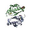
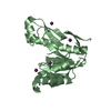
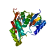
| Title | Crystal structure at 1.5 Angstroms resolution of the wild type thioredoxin-like [2Fe-2S] ferredoxin from Aquifex aeolicus | ||||||
 Components Components | [2Fe-2S] ferredoxin | ||||||
 Keywords Keywords | ELECTRON TRANSPORT / ferredoxin / thioredoxin-like fold / [2Fe-2S] cluster | ||||||
| Function / homology |  Function and homology information Function and homology information | ||||||
| Biological species |   Aquifex aeolicus (bacteria) Aquifex aeolicus (bacteria) | ||||||
| Method |  X-RAY DIFFRACTION / SYNCHROTRON / FOURIER SYNTHESIS / Resolution: 1.5 Å X-RAY DIFFRACTION / SYNCHROTRON / FOURIER SYNTHESIS / Resolution: 1.5 Å | ||||||
 Authors Authors | Yeh, A.P. / Ambroggio, X.I. / Andrade, S.L.A. / Einsle, O. / Chatelet, C. / Meyer, J. / Rees, D.C. | ||||||
 Citation Citation | Journal: J.Biol.Chem. / Year: 2002 Title: High-resolution crystal structures of the wild type and Cys-55-->Ser and Cys-59-->Ser variants of the thioredoxin-like [2Fe-2S] ferredoxin from Aquifex aeolicus Authors: Yeh, A.P. / Ambroggio, X.I. / Andrade, S.L.A. / Einsle, O. / Chatelet, C. / Meyer, J. / Rees, D.C. #1: Journal: J.Mol.Biol. / Year: 2000Title: Structure of a thioredoxin-like [2Fe-2S] ferredoxin from Aquifex aeolicus Authors: Yeh, A.P. / Chatelet, C. / Soltis, S.M. / Kuhn, P. / Meyer, J. / Rees, D.C. | ||||||
| History |
|
- Structure visualization
| Structure viewer | Molecule: MolmilJmol/JSmol |
|---|
- Downloads & links
-Download
| PDBx/mmCIF format | 1m2a.cif.gz | 59.6 KB | Display | PDBx/mmCIF format |
|---|---|---|---|---|
| PDB format | pdb1m2a.ent.gz | 43.2 KB | Display | PDB format |
| PDBx/mmJSON format | 1m2a.json.gz | Tree view | PDBx/mmJSON format | |
| Others |  Other downloads Other downloads |
-Validation report
| Arichive directory | https://data.pdbj.org/pub/pdb/validation_reports/m2/1m2aftp://data.pdbj.org/pub/pdb/validation_reports/m2/1m2a | HTTPS FTP |
|---|
-Related structure data
| Related structure data |  1m2bSC 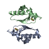 1m2dC S: Starting model for refinement C: citing same article ( |
|---|---|
| Similar structure data |


-Links
 PDBj
PDBj


- Assembly
| Deposited unit | 
| ||||||||
|---|---|---|---|---|---|---|---|---|---|
| 1 |
| ||||||||
| 2 | 
| ||||||||
| Unit cell |
| ||||||||
| Components on special symmetry positions |
|
-Components
| #1: Protein | Mass: 12206.156 Da / Num. of mol.: 2 Source method: isolated from a genetically manipulated source Source: (gene. exp.) Aquifex aeolicus (bacteria) / Gene: Fdx4 / Plasmid: pT7-7 / Production host: #2: Chemical | ChemComp-ZN /   Mass: 65.409 Da / Num. of mol.: 4 / Source method: obtained synthetically / Formula: Zn Mass: 65.409 Da / Num. of mol.: 4 / Source method: obtained synthetically / Formula: Zn#3: Chemical |   Mass: 175.820 Da / Num. of mol.: 2 / Source method: obtained synthetically / Formula: Fe2S2 Mass: 175.820 Da / Num. of mol.: 2 / Source method: obtained synthetically / Formula: Fe2S2#4: Chemical | ChemComp-SO4 / |   Mass: 96.063 Da / Num. of mol.: 1 / Source method: obtained synthetically / Formula: SO4 Mass: 96.063 Da / Num. of mol.: 1 / Source method: obtained synthetically / Formula: SO4#5: Water | ChemComp-HOH / |  Mass: 18.015 Da / Num. of mol.: 187 / Source method: isolated from a natural source / Formula: H2O Mass: 18.015 Da / Num. of mol.: 187 / Source method: isolated from a natural source / Formula: H2O |
|---|
-Experimental details
-Experiment
| Experiment | Method: X-RAY DIFFRACTION / Number of used crystals: 1 |
|---|
- Sample preparation
| Crystal | Density Matthews: 1.82 Å3/Da / Density % sol: 32.42 % | |||||||||||||||||||||||||||||||||||||||||||||||||
|---|---|---|---|---|---|---|---|---|---|---|---|---|---|---|---|---|---|---|---|---|---|---|---|---|---|---|---|---|---|---|---|---|---|---|---|---|---|---|---|---|---|---|---|---|---|---|---|---|---|---|
| Crystal grow | Temperature: 298 K / Method: vapor diffusion, sitting drop / pH: 6.5 Details: zinc sulfate heptahydrate, MES buffer, polyethylene glycol monomethyl ether 550, pH 6.5, VAPOR DIFFUSION, SITTING DROP, temperature 298K | |||||||||||||||||||||||||||||||||||||||||||||||||
| Crystal grow | *PLUS pH: 8 | |||||||||||||||||||||||||||||||||||||||||||||||||
| Components of the solutions | *PLUS
|
-Data collection
| Diffraction | Mean temperature: 100 K |
|---|---|
| Diffraction source | Source: SYNCHROTRON / Site: SSRL  / Beamline: BL9-2 / Wavelength: 0.9918 Å / Beamline: BL9-2 / Wavelength: 0.9918 Å |
| Detector | Type: ADSC QUANTUM 315 / Detector: CCD / Date: Feb 18, 2002 |
| Radiation | Monochromator: graphite / Protocol: SINGLE WAVELENGTH / Monochromatic (M) / Laue (L): M / Scattering type: x-ray |
| Radiation wavelength | Wavelength: 0.9918 Å / Relative weight: 1 |
| Reflection | Resolution: 1.5→31.5 Å / Num. all: 27758 / Num. obs: 27758 / % possible obs: 98.9 % / Observed criterion σ(F): 1 / Observed criterion σ(I): 2 / Redundancy: 3.6 % / Rmerge(I) obs: 0.051 / Rsym value: 0.051 / Net I/σ(I): 7.8 |
| Reflection shell | Resolution: 1.5→1.54 Å / Redundancy: 3.3 % / Rmerge(I) obs: 0.404 / Mean I/σ(I) obs: 1.5 / Num. unique all: 1969 / Rsym value: 0.404 / % possible all: 97.1 |
| Reflection | *PLUS Highest resolution: 1.5 Å / Num. measured all: 98554 / Rmerge(I) obs: 0.051 |
| Reflection shell | *PLUS % possible obs: 97.1 % / Rmerge(I) obs: 0.404 |
- Processing
| Software |
| |||||||||||||||||||||||||
|---|---|---|---|---|---|---|---|---|---|---|---|---|---|---|---|---|---|---|---|---|---|---|---|---|---|---|
| Refinement | Method to determine structure: FOURIER SYNTHESIS Starting model: PDB ENTRY 1M2B, Cys55Ser Aquifex aeolicus [2Fe-2S] ferredoxin structure Resolution: 1.5→31.5 Å / σ(F): 0 / Stereochemistry target values: Engh & Huber
| |||||||||||||||||||||||||
| Refine analyze | Luzzati coordinate error obs: 0.16 Å | |||||||||||||||||||||||||
| Refinement step | Cycle: LAST / Resolution: 1.5→31.5 Å
| |||||||||||||||||||||||||
| Refine LS restraints |
| |||||||||||||||||||||||||
| Refinement | *PLUS Highest resolution: 1.5 Å / % reflection Rfree: 3 % / Rfactor Rfree: 0.216 / Rfactor Rwork: 0.184 | |||||||||||||||||||||||||
| Solvent computation | *PLUS | |||||||||||||||||||||||||
| Displacement parameters | *PLUS | |||||||||||||||||||||||||
| Refine LS restraints | *PLUS
|
