 Movie
Movie Controller

[English] 日本語
 Yorodumi
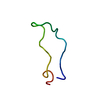
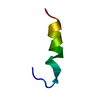
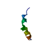
- PDB-1m25: STRUCTURE OF SYNTHETIC 26-MER PEPTIDE CONTAINING 145-169 SHEEP PR... -
+ Open data

- Basic information
| Entry | Database: PDB / ID: 1m25 | ||||||
|---|---|---|---|---|---|---|---|
| Title | STRUCTURE OF SYNTHETIC 26-MER PEPTIDE CONTAINING 145-169 SHEEP PRION PROTEIN SEGMENT AND C-TERMINAL CYSTEINE IN TFE SOLUTION | ||||||
 Components Components | MAJOR PRION PROTEIN | ||||||
 Keywords Keywords | UNKNOWN FUNCTION / helix / prion / TFE | ||||||
| Function / homology |  Function and homology information Function and homology informationside of membrane / tubulin binding / protein homooligomerization / microtubule binding / copper ion binding / Golgi apparatus / identical protein binding / plasma membrane Similarity search - Function | ||||||
| Method | SOLUTION NMR / simulated annealing using torsion angle dynamics as implemented in the program CNS | ||||||
 Authors Authors | Megy, S. / Bertho, G. / Kozin, S.A. / Coadou, G. / Debey, P. / Hoa, G.H. / Girault, J.-P. | ||||||
 Citation Citation | Journal: Protein Sci. / Year: 2004 Title: Possible role of region 152-156 in the structural duality of a peptide fragment from sheep prion protein Authors: Megy, S. / Bertho, G. / Kozin, S.A. / Debey, P. / Hoa, G.H. / Girault, J.-P. #1: Journal: J.Biol.Chem. / Year: 2001Title: Sheep prion protein synthetic peptide spanning helix 1 and beta-strand 2 (residues 142-166) shows beta-hairpin structure in solution Authors: Kozin, S.A. / Bertho, G. / Mazur, A.K. / Rabesona, H. / Girault, J.-P. / Haertle, T. / Takahashi, M. / Debey, P. / Hoa, G.H. | ||||||
| History |
|
- Structure visualization
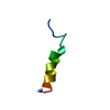
| Structure viewer | Molecule: MolmilJmol/JSmol |
|---|
- Downloads & links
-Download
| PDBx/mmCIF format | 1m25.cif.gz | 182.6 KB | Display | PDBx/mmCIF format |
|---|---|---|---|---|
| PDB format | pdb1m25.ent.gz | 150.8 KB | Display | PDB format |
| PDBx/mmJSON format | 1m25.json.gz | Tree view | PDBx/mmJSON format | |
| Others |  Other downloads Other downloads |
-Validation report
| Arichive directory | https://data.pdbj.org/pub/pdb/validation_reports/m2/1m25ftp://data.pdbj.org/pub/pdb/validation_reports/m2/1m25 | HTTPS FTP |
|---|
-Related structure data
| Related structure data | |
|---|---|
| Similar structure data | |
| Other databases |
|


-Links
 PDBj
PDBj

- Assembly
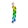
| Deposited unit | 
| |||||||||
|---|---|---|---|---|---|---|---|---|---|---|
| 1 |
| |||||||||
| NMR ensembles |
|
-Components
| #1: Protein/peptide | Mass: 3431.729 Da / Num. of mol.: 1 / Fragment: Residues 145-169 / Source method: obtained synthetically Details: THIS PEPTIDE WAS CHEMICALLY SYNTHESIZED. EXCEPT FOR THE C-TERMINAL CYSTEINE, THIS SEQUENCE OCCURS NATURALLY IN OVIS ARIES (SHEEP). References: UniProt: P23907 |
|---|
-Experimental details
-Experiment
| Experiment | Method: SOLUTION NMR |
|---|---|
| NMR experiment | Type: 2D NOESY |
| NMR details | Text: This structure was determined using standard 2D homonuclear techniques. |
- Sample preparation
| Details | Contents: 4.5mM Solvent system: 87/13 (v/v), CF3CD2OH / 10mM sodium phosphate buffer, pH 6.5 |
|---|---|
| Sample conditions | Ionic strength: 1mM / pH: 6.5 / Pressure: 1 atm / Temperature: 278 K |
| Crystal grow | *PLUS Method: other / Details: NMR |
-NMR measurement
| Radiation | Protocol: SINGLE WAVELENGTH / Monochromatic (M) / Laue (L): M |
|---|---|
| Radiation wavelength | Relative weight: 1 |
| NMR spectrometer | Type: Bruker AMX / Manufacturer: Bruker / Model: AMX / Field strength: 500 MHz |
- Processing
| NMR software |
| ||||||||||||||||
|---|---|---|---|---|---|---|---|---|---|---|---|---|---|---|---|---|---|
| Refinement | Method: simulated annealing using torsion angle dynamics as implemented in the program CNS Software ordinal: 1 Details: the structures are based on a total of 252 interresidual NOE-derived distance constraints, 22 dihedral angle restraints, 25 CSI restraints from Ha, Ca, Cb. | ||||||||||||||||
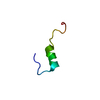
| NMR representative | Selection criteria: lowest energy, without noe violations | ||||||||||||||||
| NMR ensemble | Conformer selection criteria: back calculated data agree with experimental NOESY spectrum,structures with the lowest energy Conformers calculated total number: 200 / Conformers submitted total number: 20 |
