+ データを開く
- 基本情報
基本情報
| 登録情報 | データベース: PDB / ID: 1m11 | ||||||
|---|---|---|---|---|---|---|---|
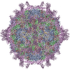
| タイトル | structural model of human decay-accelerating factor bound to echovirus 7 from cryo-electron microscopy | ||||||
 要素 要素 |
| ||||||
 キーワード キーワード | Virus/Receptor / decay-accelerating factor / SCR / Icosahedral virus / Virus-Receptor COMPLEX | ||||||
| 機能・相同性 |  機能・相同性情報 機能・相同性情報regulation of lipopolysaccharide-mediated signaling pathway / negative regulation of complement activation / regulation of complement-dependent cytotoxicity / regulation of complement activation / respiratory burst / positive regulation of CD4-positive, alpha-beta T cell activation / positive regulation of CD4-positive, alpha-beta T cell proliferation / Class B/2 (Secretin family receptors) / symbiont-mediated suppression of host cytoplasmic pattern recognition receptor signaling pathway via inhibition of RIG-I activity / ficolin-1-rich granule membrane ...regulation of lipopolysaccharide-mediated signaling pathway / negative regulation of complement activation / regulation of complement-dependent cytotoxicity / regulation of complement activation / respiratory burst / positive regulation of CD4-positive, alpha-beta T cell activation / positive regulation of CD4-positive, alpha-beta T cell proliferation / Class B/2 (Secretin family receptors) / symbiont-mediated suppression of host cytoplasmic pattern recognition receptor signaling pathway via inhibition of RIG-I activity / ficolin-1-rich granule membrane / complement activation, classical pathway / transport vesicle / side of membrane / COPI-mediated anterograde transport / endoplasmic reticulum-Golgi intermediate compartment membrane / ribonucleoside triphosphate phosphatase activity / secretory granule membrane / Regulation of Complement cascade / picornain 2A / symbiont-mediated suppression of host mRNA export from nucleus / symbiont genome entry into host cell via pore formation in plasma membrane / picornain 3C / T=pseudo3 icosahedral viral capsid / positive regulation of T cell cytokine production / host cell cytoplasmic vesicle membrane / nucleoside-triphosphate phosphatase / channel activity / positive regulation of cytosolic calcium ion concentration / virus receptor activity / monoatomic ion transmembrane transport / DNA replication / RNA helicase activity / membrane raft / endocytosis involved in viral entry into host cell / Golgi membrane / symbiont-mediated activation of host autophagy / innate immune response / RNA-directed RNA polymerase / cysteine-type endopeptidase activity / viral RNA genome replication / RNA-directed RNA polymerase activity / DNA-templated transcription / lipid binding / Neutrophil degranulation / virion attachment to host cell / host cell nucleus / structural molecule activity / cell surface / proteolysis / RNA binding / extracellular exosome / extracellular region / zinc ion binding / ATP binding / membrane / plasma membrane 類似検索 - 分子機能 | ||||||
| 生物種 |  Homo sapiens (ヒト) Homo sapiens (ヒト) Human echovirus 7 (ウイルス) Human echovirus 7 (ウイルス) | ||||||
| 手法 | 電子顕微鏡法 / 単粒子再構成法 / クライオ電子顕微鏡法 / 解像度: 16 Å | ||||||
 データ登録者 データ登録者 | He, Y. / Lin, F. / Chipman, P.R. / Bator, C.M. / Baker, T.S. / Shoham, M. / Kuhn, R.J. / Medof, M.E. / Rossmann, M.G. | ||||||
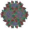
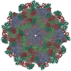
 引用 引用 |  ジャーナル: Proc Natl Acad Sci U S A / 年: 2002 ジャーナル: Proc Natl Acad Sci U S A / 年: 2002タイトル: Structure of decay-accelerating factor bound to echovirus 7: a virus-receptor complex. 著者: Yongning He / Feng Lin / Paul R Chipman / Carol M Bator / Timothy S Baker / Menachem Shoham / Richard J Kuhn / M Edward Medof / Michael G Rossmann /  要旨: Echoviruses are enteroviruses that belong to Picornaviridae. Many echoviruses use decay-accelerating factor (DAF) as their cellular receptor. DAF is a glycosylphosphatidyl inositol-anchored ...Echoviruses are enteroviruses that belong to Picornaviridae. Many echoviruses use decay-accelerating factor (DAF) as their cellular receptor. DAF is a glycosylphosphatidyl inositol-anchored complement regulatory protein found on most cell surfaces. It functions to protect cells from complement attack. The cryo-electron microscopy reconstructions of echovirus 7 complexed with DAF show that the DAF-binding regions are located close to the icosahedral twofold axes, in contrast to other enterovirus complexes where the viral canyon is the receptor binding site. This novel receptor binding position suggests that DAF is important for the attachment of viral particles to host cells, but probably not for initiating viral uncoating, as is the case with canyon-binding receptors. Thus, a different cell entry mechanism must be used for enteroviruses that bind DAF. | ||||||
| 履歴 |
|
- 構造の表示
構造の表示
| ムービー |
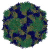
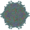
 ムービービューア ムービービューア |
|---|---|
| 構造ビューア | 分子:  Molmil Molmil Jmol/JSmol Jmol/JSmol |
- ダウンロードとリンク
- ダウンロード
| PDBx/mmCIF形式 |  1m11.cif.gz 1m11.cif.gz | 46.6 KB | 表示 |  PDBx/mmCIF形式 PDBx/mmCIF形式 |
|---|---|---|---|---|
| PDB形式 |  pdb1m11.ent.gz pdb1m11.ent.gz | 23.8 KB | 表示 |  PDB形式 PDB形式 |
| PDBx/mmJSON形式 |  1m11.json.gz 1m11.json.gz | ツリー表示 |  PDBx/mmJSON形式 PDBx/mmJSON形式 | |
| その他 |  その他のダウンロード その他のダウンロード |
-検証レポート
| 文書・要旨 |  1m11_validation.pdf.gz 1m11_validation.pdf.gz | 282.6 KB | 表示 |  wwPDB検証レポート wwPDB検証レポート |
|---|---|---|---|---|
| 文書・詳細版 |  1m11_full_validation.pdf.gz 1m11_full_validation.pdf.gz | 282.2 KB | 表示 | |
| XML形式データ |  1m11_validation.xml.gz 1m11_validation.xml.gz | 903 B | 表示 | |
| CIF形式データ |  1m11_validation.cif.gz 1m11_validation.cif.gz | 10.1 KB | 表示 | |
| アーカイブディレクトリ |  https://data.pdbj.org/pub/pdb/validation_reports/m1/1m11 https://data.pdbj.org/pub/pdb/validation_reports/m1/1m11 ftp://data.pdbj.org/pub/pdb/validation_reports/m1/1m11 ftp://data.pdbj.org/pub/pdb/validation_reports/m1/1m11 | HTTPS FTP |
-関連構造データ
| 関連構造データ |  1g40 |
|---|---|
| 類似構造データ |
- リンク
リンク
- 集合体
集合体
| 登録構造単位 | 
|
|---|---|
| 1 | x 60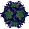
|
| 2 |
|
| 3 | x 5
|
| 4 | x 6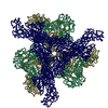
|
| 5 | 
|
| 対称性 | 点対称性: (ヘルマン・モーガン記号: 532 / シェーンフリース記号: I (正20面体型対称)) |
- 要素
要素
| #1: タンパク質 | 分子量: 27017.299 Da / 分子数: 1 / 断片: four SCR domains 1 to 4 / 由来タイプ: 組換発現 / 由来: (組換発現)  Homo sapiens (ヒト) / 発現宿主: Homo sapiens (ヒト) / 発現宿主:  Pichia pastoris (菌類) / 参照: UniProt: P08174 Pichia pastoris (菌類) / 参照: UniProt: P08174 |
|---|---|
| #2: タンパク質 | 分子量: 31682.414 Da / 分子数: 1 / 由来タイプ: 組換発現 / 由来: (組換発現)  Human echovirus 7 (ウイルス) / 属: Enterovirus / 生物種: Human enterovirus B / 解説: RHABDOMYOSARCOMA CELL (RD); / 細胞株 (発現宿主): RD / 発現宿主: Human echovirus 7 (ウイルス) / 属: Enterovirus / 生物種: Human enterovirus B / 解説: RHABDOMYOSARCOMA CELL (RD); / 細胞株 (発現宿主): RD / 発現宿主:  Homo sapiens (ヒト) / 組織 (発現宿主): muscle / 参照: UniProt: Q914E0 Homo sapiens (ヒト) / 組織 (発現宿主): muscle / 参照: UniProt: Q914E0 |
| #3: タンパク質 | 分子量: 28443.154 Da / 分子数: 1 / 由来タイプ: 組換発現 / 由来: (組換発現)  Human echovirus 7 (ウイルス) / 属: Enterovirus / 生物種: Human enterovirus B / 解説: RHABDOMYOSARCOMA CELL (RD); / 細胞株 (発現宿主): RD / 発現宿主: Human echovirus 7 (ウイルス) / 属: Enterovirus / 生物種: Human enterovirus B / 解説: RHABDOMYOSARCOMA CELL (RD); / 細胞株 (発現宿主): RD / 発現宿主:  Homo sapiens (ヒト) / 組織 (発現宿主): muscle / 参照: UniProt: Q914E0 Homo sapiens (ヒト) / 組織 (発現宿主): muscle / 参照: UniProt: Q914E0 |
| #4: タンパク質 | 分子量: 26264.000 Da / 分子数: 1 / 由来タイプ: 組換発現 / 由来: (組換発現)  Human echovirus 7 (ウイルス) / 属: Enterovirus / 生物種: Human enterovirus B / 解説: RHABDOMYOSARCOMA CELL (RD) / 細胞株 (発現宿主): RD / 発現宿主: Human echovirus 7 (ウイルス) / 属: Enterovirus / 生物種: Human enterovirus B / 解説: RHABDOMYOSARCOMA CELL (RD) / 細胞株 (発現宿主): RD / 発現宿主:  Homo sapiens (ヒト) / 組織 (発現宿主): muscle / 参照: UniProt: Q914E0 Homo sapiens (ヒト) / 組織 (発現宿主): muscle / 参照: UniProt: Q914E0 |
-実験情報
-実験
| 実験 | 手法: 電子顕微鏡法 |
|---|---|
| EM実験 | 試料の集合状態: PARTICLE / 3次元再構成法: 単粒子再構成法 |
- 試料調製
試料調製
| 構成要素 | 名称: human decay-accelerating factor, HUMAN ECHOVIRUS 7 COAT PROTEINS タイプ: VIRUS 詳細: This structure is modeled based on cryo-EM density at 16A resolution. |
|---|---|
| 緩衝液 | 名称: tris buffer pH7.5 / pH: 7.5 / 詳細: tris buffer pH7.5 |
| 試料 | 濃度: 8 mg/ml / 包埋: NO / シャドウイング: NO / 染色: NO / 凍結: YES |
| 急速凍結 | 詳細: SAMPLES WERE PREPARED AS THIN LAYERS OF VITREOUS ICE AND MAINTAINED AT NEAR LIQUID NITROGEN TEMPERATURE IN THE ELECTRON MICROSCOPE WITH A GATAN 626 CRYOTRANSFER HOLDER |
| 結晶化 | *PLUS 手法: cryo-electron microscopy |
- 電子顕微鏡撮影
| 顕微鏡 | モデル: FEI/PHILIPS CM300FEG/T / 日付: 2001年9月10日 |
|---|---|
| 電子銃 | 電子線源:  FIELD EMISSION GUN / 加速電圧: 300 kV / 照射モード: FLOOD BEAM FIELD EMISSION GUN / 加速電圧: 300 kV / 照射モード: FLOOD BEAM |
| 電子レンズ | モード: BRIGHT FIELD / 倍率(公称値): 45000 X / 最大 デフォーカス(公称値): 4200 nm / 最小 デフォーカス(公称値): 1800 nm |
| 試料ホルダ | 温度: 120 K |
| 撮影 | 電子線照射量: 16.6 e/Å2 |
- 解析
解析
| EMソフトウェア |
| ||||||||||||||||||||||||||||
|---|---|---|---|---|---|---|---|---|---|---|---|---|---|---|---|---|---|---|---|---|---|---|---|---|---|---|---|---|---|
| CTF補正 | 詳細: CTF correction of each micrograph | ||||||||||||||||||||||||||||
| 対称性 | 点対称性: I (正20面体型対称) | ||||||||||||||||||||||||||||
| 3次元再構成 | 手法: PFT / 解像度: 16 Å / ピクセルサイズ(公称値): 3.11 Å 詳細: The echovirus 7 structure is unknown, the model used here is from coxsackievirus B3 (1COV) and echovirus 1 (1EV1).The DAF receptor model is from 1g40. Only CA coordinates are presented in the entry. 対称性のタイプ: POINT | ||||||||||||||||||||||||||||
| 原子モデル構築 | 空間: REAL | ||||||||||||||||||||||||||||
| 原子モデル構築 |
| ||||||||||||||||||||||||||||
| 精密化 | 最高解像度: 16 Å | ||||||||||||||||||||||||||||
| 精密化ステップ | サイクル: LAST / 最高解像度: 16 Å
|
 ムービー
ムービー コントローラー






 PDBj
PDBj


