 Movie
Movie Controller

[English] 日本語
 Yorodumi
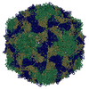
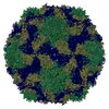
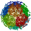
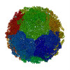
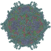
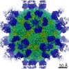
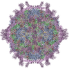
- PDB-1m11: structural model of human decay-accelerating factor bound to echo... -
+ Open data

- Basic information
| Entry | Database: PDB / ID: 1m11 | ||||||
|---|---|---|---|---|---|---|---|
| Title | structural model of human decay-accelerating factor bound to echovirus 7 from cryo-electron microscopy | ||||||
 Components Components |
| ||||||
 Keywords Keywords | Virus/Receptor / decay-accelerating factor / SCR / Icosahedral virus / Virus-Receptor COMPLEX | ||||||
| Function / homology |  Function and homology information Function and homology informationregulation of lipopolysaccharide-mediated signaling pathway / negative regulation of complement activation / regulation of complement-dependent cytotoxicity / regulation of complement activation / positive regulation of CD4-positive, alpha-beta T cell activation / respiratory burst / positive regulation of CD4-positive, alpha-beta T cell proliferation / Class B/2 (Secretin family receptors) / symbiont-mediated suppression of host cytoplasmic pattern recognition receptor signaling pathway via inhibition of RIG-I activity / ficolin-1-rich granule membrane ...regulation of lipopolysaccharide-mediated signaling pathway / negative regulation of complement activation / regulation of complement-dependent cytotoxicity / regulation of complement activation / positive regulation of CD4-positive, alpha-beta T cell activation / respiratory burst / positive regulation of CD4-positive, alpha-beta T cell proliferation / Class B/2 (Secretin family receptors) / symbiont-mediated suppression of host cytoplasmic pattern recognition receptor signaling pathway via inhibition of RIG-I activity / ficolin-1-rich granule membrane / complement activation, classical pathway / side of membrane / transport vesicle / COPI-mediated anterograde transport / endoplasmic reticulum-Golgi intermediate compartment membrane / Regulation of Complement cascade / secretory granule membrane / picornain 2A / symbiont-mediated suppression of host mRNA export from nucleus / symbiont genome entry into host cell via pore formation in plasma membrane / picornain 3C / T=pseudo3 icosahedral viral capsid / positive regulation of T cell cytokine production / host cell cytoplasmic vesicle membrane / ribonucleoside triphosphate phosphatase activity / nucleoside-triphosphate phosphatase / positive regulation of cytosolic calcium ion concentration / virus receptor activity / channel activity / monoatomic ion transmembrane transport / DNA replication / RNA helicase activity / endocytosis involved in viral entry into host cell / membrane raft / Golgi membrane / symbiont-mediated activation of host autophagy / innate immune response / RNA-directed RNA polymerase / cysteine-type endopeptidase activity / viral RNA genome replication / RNA-directed RNA polymerase activity / Neutrophil degranulation / lipid binding / virion attachment to host cell / host cell nucleus / structural molecule activity / cell surface / DNA-templated transcription / proteolysis / RNA binding / extracellular exosome / extracellular region / zinc ion binding / ATP binding / plasma membrane Similarity search - Function | ||||||
| Biological species |  Homo sapiens (human) Homo sapiens (human) Human echovirus 7 Human echovirus 7 | ||||||
| Method | ELECTRON MICROSCOPY / single particle reconstruction / cryo EM / Resolution: 16 Å | ||||||
 Authors Authors | He, Y. / Lin, F. / Chipman, P.R. / Bator, C.M. / Baker, T.S. / Shoham, M. / Kuhn, R.J. / Medof, M.E. / Rossmann, M.G. | ||||||
 Citation Citation | Journal: Proc Natl Acad Sci U S A / Year: 2002 Title: Structure of decay-accelerating factor bound to echovirus 7: a virus-receptor complex. Authors: Yongning He / Feng Lin / Paul R Chipman / Carol M Bator / Timothy S Baker / Menachem Shoham / Richard J Kuhn / M Edward Medof / Michael G Rossmann /  Abstract: Echoviruses are enteroviruses that belong to Picornaviridae. Many echoviruses use decay-accelerating factor (DAF) as their cellular receptor. DAF is a glycosylphosphatidyl inositol-anchored ...Echoviruses are enteroviruses that belong to Picornaviridae. Many echoviruses use decay-accelerating factor (DAF) as their cellular receptor. DAF is a glycosylphosphatidyl inositol-anchored complement regulatory protein found on most cell surfaces. It functions to protect cells from complement attack. The cryo-electron microscopy reconstructions of echovirus 7 complexed with DAF show that the DAF-binding regions are located close to the icosahedral twofold axes, in contrast to other enterovirus complexes where the viral canyon is the receptor binding site. This novel receptor binding position suggests that DAF is important for the attachment of viral particles to host cells, but probably not for initiating viral uncoating, as is the case with canyon-binding receptors. Thus, a different cell entry mechanism must be used for enteroviruses that bind DAF. | ||||||
| History |
|
- Structure visualization
| Movie |
Movie viewer |
|---|---|
| Structure viewer | Molecule: MolmilJmol/JSmol |
- Downloads & links
-Download
| PDBx/mmCIF format | 1m11.cif.gz | 46.6 KB | Display | PDBx/mmCIF format |
|---|---|---|---|---|
| PDB format | pdb1m11.ent.gz | 23.8 KB | Display | PDB format |
| PDBx/mmJSON format | 1m11.json.gz | Tree view | PDBx/mmJSON format | |
| Others |  Other downloads Other downloads |
-Validation report
| Arichive directory | https://data.pdbj.org/pub/pdb/validation_reports/m1/1m11ftp://data.pdbj.org/pub/pdb/validation_reports/m1/1m11 | HTTPS FTP |
|---|
-Related structure data
| Related structure data |  1g40 |
|---|---|
| Similar structure data |
-Links
 PDBj
PDBj


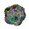
- Assembly
| Deposited unit | 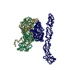
|
|---|---|
| 1 | x 60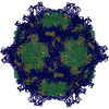
|
| 2 |
|
| 3 | x 5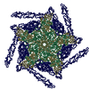
|
| 4 | x 6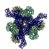
|
| 5 | 
|
| Symmetry | Point symmetry: (Hermann–Mauguin notation: 532 / Schoenflies symbol: I (icosahedral)) |
-Components
| #1: Protein | Mass: 27017.299 Da / Num. of mol.: 1 / Fragment: four SCR domains 1 to 4 Source method: isolated from a genetically manipulated source Source: (gene. exp.) Homo sapiens (human) / Production host:  Pichia pastoris (fungus) / References: UniProt: P08174 Pichia pastoris (fungus) / References: UniProt: P08174 |
|---|---|
| #2: Protein | Mass: 31682.414 Da / Num. of mol.: 1 Source method: isolated from a genetically manipulated source Source: (gene. exp.) Human echovirus 7 / Genus: Enterovirus / Species: Human enterovirus B / Description: RHABDOMYOSARCOMA CELL (RD); / Cell line (production host): RD / Production host: Homo sapiens (human) / Tissue (production host): muscle / References: UniProt: Q914E0 |
| #3: Protein | Mass: 28443.154 Da / Num. of mol.: 1 Source method: isolated from a genetically manipulated source Source: (gene. exp.) Human echovirus 7 / Genus: Enterovirus / Species: Human enterovirus B / Description: RHABDOMYOSARCOMA CELL (RD); / Cell line (production host): RD / Production host: Homo sapiens (human) / Tissue (production host): muscle / References: UniProt: Q914E0 |
| #4: Protein | Mass: 26264.000 Da / Num. of mol.: 1 Source method: isolated from a genetically manipulated source Source: (gene. exp.) Human echovirus 7 / Genus: Enterovirus / Species: Human enterovirus B / Description: RHABDOMYOSARCOMA CELL (RD) / Cell line (production host): RD / Production host: Homo sapiens (human) / Tissue (production host): muscle / References: UniProt: Q914E0 |
-Experimental details
-Experiment
| Experiment | Method: ELECTRON MICROSCOPY |
|---|---|
| EM experiment | Aggregation state: PARTICLE / 3D reconstruction method: single particle reconstruction |
- Sample preparation
| Component | Name: human decay-accelerating factor, HUMAN ECHOVIRUS 7 COAT PROTEINS Type: VIRUS Details: This structure is modeled based on cryo-EM density at 16A resolution. |
|---|---|
| Buffer solution | Name: tris buffer pH7.5 / pH: 7.5 / Details: tris buffer pH7.5 |
| Specimen | Conc.: 8 mg/ml / Embedding applied: NO / Shadowing applied: NO / Staining applied: NO / Vitrification applied: YES |
| Vitrification | Details: SAMPLES WERE PREPARED AS THIN LAYERS OF VITREOUS ICE AND MAINTAINED AT NEAR LIQUID NITROGEN TEMPERATURE IN THE ELECTRON MICROSCOPE WITH A GATAN 626 CRYOTRANSFER HOLDER |
| Crystal grow | *PLUS Method: cryo-electron microscopy |
- Electron microscopy imaging
| Microscopy | Model: FEI/PHILIPS CM300FEG/T / Date: Sep 10, 2001 |
|---|---|
| Electron gun | Electron source:  FIELD EMISSION GUN / Accelerating voltage: 300 kV / Illumination mode: FLOOD BEAM FIELD EMISSION GUN / Accelerating voltage: 300 kV / Illumination mode: FLOOD BEAM |
| Electron lens | Mode: BRIGHT FIELD / Nominal magnification: 45000 X / Nominal defocus max: 4200 nm / Nominal defocus min: 1800 nm |
| Specimen holder | Temperature: 120 K |
| Image recording | Electron dose: 16.6 e/Å2 |
- Processing
| EM software |
| ||||||||||||||||||||||||||||
|---|---|---|---|---|---|---|---|---|---|---|---|---|---|---|---|---|---|---|---|---|---|---|---|---|---|---|---|---|---|
| CTF correction | Details: CTF correction of each micrograph | ||||||||||||||||||||||||||||
| Symmetry | Point symmetry: I (icosahedral) | ||||||||||||||||||||||||||||
| 3D reconstruction | Method: PFT / Resolution: 16 Å / Nominal pixel size: 3.11 Å Details: The echovirus 7 structure is unknown, the model used here is from coxsackievirus B3 (1COV) and echovirus 1 (1EV1).The DAF receptor model is from 1g40. Only CA coordinates are presented in the entry. Symmetry type: POINT | ||||||||||||||||||||||||||||
| Atomic model building | Space: REAL | ||||||||||||||||||||||||||||
| Atomic model building |
| ||||||||||||||||||||||||||||
| Refinement | Highest resolution: 16 Å | ||||||||||||||||||||||||||||
| Refinement step | Cycle: LAST / Highest resolution: 16 Å
|
