 Movie
Movie Controller

[English] 日本語
 Yorodumi
- PDB-1lzv: Site-Specific Mutant (Tyr7 replaced with His) of Human Carbonic A... -
+ Open data

- Basic information
| Entry | Database: PDB / ID: 1lzv | ||||||
|---|---|---|---|---|---|---|---|
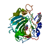
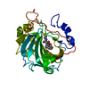
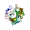
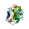
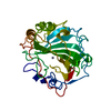
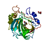
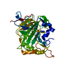
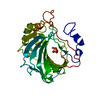
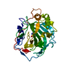
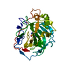
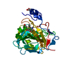
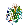
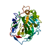
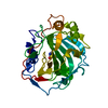
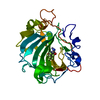
| Title | Site-Specific Mutant (Tyr7 replaced with His) of Human Carbonic Anhydrase II | ||||||
 Components Components | Carbonic Anhydrase II | ||||||
 Keywords Keywords | LYASE / Twisted Beta Sheet / Zinc Metalloenzyme | ||||||
| Function / homology |  Function and homology information Function and homology informationpositive regulation of dipeptide transmembrane transport / : / regulation of monoatomic anion transport / secretion / cyanamide hydratase / cyanamide hydratase activity / arylesterase activity / regulation of chloride transport / Reversible hydration of carbon dioxide / morphogenesis of an epithelium ...positive regulation of dipeptide transmembrane transport / : / regulation of monoatomic anion transport / secretion / cyanamide hydratase / cyanamide hydratase activity / arylesterase activity / regulation of chloride transport / Reversible hydration of carbon dioxide / morphogenesis of an epithelium / angiotensin-activated signaling pathway / Developmental Lineage of Pancreatic Ductal Cells / carbonic anhydrase / regulation of intracellular pH / carbonate dehydratase activity / positive regulation of synaptic transmission, GABAergic / carbon dioxide transport / Erythrocytes take up oxygen and release carbon dioxide / Erythrocytes take up carbon dioxide and release oxygen / neuron cellular homeostasis / apical part of cell / myelin sheath / extracellular exosome / zinc ion binding / plasma membrane / cytoplasm / cytosol Similarity search - Function | ||||||
| Biological species |  Homo sapiens (human) Homo sapiens (human) | ||||||
| Method |  X-RAY DIFFRACTION / MOLECULAR REPLACEMENT / Resolution: 2.3 Å X-RAY DIFFRACTION / MOLECULAR REPLACEMENT / Resolution: 2.3 Å | ||||||
 Authors Authors | Tu, C.K. / Qian, M. / An, H. / Wadhwa, N.R. / Duda, D.M. / Yoshioka, C. / Pathak, Y. / McKenna, R. / Laipis, P.J. / Silverman, D.N. | ||||||
 Citation Citation | Journal: J.Biol.Chem. / Year: 2002 Title: Kinetic analysis of multiple proton shuttles in the active site of human carbonic anhydrase. Authors: Tu, C. / Qian, M. / An, H. / Wadhwa, N.R. / Duda, D. / Yoshioka, C. / Pathak, Y. / McKenna, R. / Laipis, P.J. / Silverman, D.N. | ||||||
| History |
| ||||||
| Remark 999 | SEQUENCE Residue 125 is covalently bound to 127. Number 126 is not used as a matter of convention ...SEQUENCE Residue 125 is covalently bound to 127. Number 126 is not used as a matter of convention for sequence alignments. |
- Structure visualization
| Structure viewer | Molecule: MolmilJmol/JSmol |
|---|
- Downloads & links
-Download
| PDBx/mmCIF format | 1lzv.cif.gz | 64.3 KB | Display | PDBx/mmCIF format |
|---|---|---|---|---|
| PDB format | pdb1lzv.ent.gz | 46.5 KB | Display | PDB format |
| PDBx/mmJSON format | 1lzv.json.gz | Tree view | PDBx/mmJSON format | |
| Others |  Other downloads Other downloads |
-Validation report
| Arichive directory | https://data.pdbj.org/pub/pdb/validation_reports/lz/1lzvftp://data.pdbj.org/pub/pdb/validation_reports/lz/1lzv | HTTPS FTP |
|---|
-Related structure data
| Related structure data | 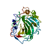 1g0fS S: Starting model for refinement |
|---|---|
| Similar structure data |


-Links
 PDBj
PDBj

- Assembly
| Deposited unit | 
| ||||||||
|---|---|---|---|---|---|---|---|---|---|
| 1 |
| ||||||||
| Unit cell |
| ||||||||
| Details | The Biological Assembly is a Monomer Constructed from Chain A |
-Components
| #1: Protein | Mass: 29264.035 Da / Num. of mol.: 1 / Mutation: Y7H Source method: isolated from a genetically manipulated source Source: (gene. exp.) Homo sapiens (human) / Production host:  |
|---|---|
| #2: Chemical | ChemComp-ZN /   Mass: 65.409 Da / Num. of mol.: 1 / Source method: obtained synthetically / Formula: Zn Mass: 65.409 Da / Num. of mol.: 1 / Source method: obtained synthetically / Formula: Zn |
| #3: Water | ChemComp-HOH /  Mass: 18.015 Da / Num. of mol.: 56 / Source method: isolated from a natural source / Formula: H2O Mass: 18.015 Da / Num. of mol.: 56 / Source method: isolated from a natural source / Formula: H2O |
-Experimental details
-Experiment
| Experiment | Method: X-RAY DIFFRACTION / Number of used crystals: 3 |
|---|
- Sample preparation
| Crystal | Density Matthews: 2.15 Å3/Da / Density % sol: 42.73 % | ||||||||||||||||||||||||||||||
|---|---|---|---|---|---|---|---|---|---|---|---|---|---|---|---|---|---|---|---|---|---|---|---|---|---|---|---|---|---|---|---|
| Crystal grow | Temperature: 277 K / Method: vapor diffusion, hanging drop / pH: 7.8 Details: Ammonium Sulfate, Tris HCL, pH 7.8, VAPOR DIFFUSION, HANGING DROP, temperature 277K | ||||||||||||||||||||||||||||||
| Crystal grow | *PLUS Temperature: 40 ℃ | ||||||||||||||||||||||||||||||
| Components of the solutions | *PLUS
|
-Data collection
| Diffraction | Mean temperature: 293 K |
|---|---|
| Diffraction source | Source: ROTATING ANODE / Type: RIGAKU RU300 / Wavelength: 1.548 Å |
| Detector | Type: RIGAKU RAXIS IV / Detector: IMAGE PLATE / Date: Apr 10, 2002 |
| Radiation | Monochromator: Osmic Optics / Protocol: SINGLE WAVELENGTH / Monochromatic (M) / Laue (L): M / Scattering type: x-ray |
| Radiation wavelength | Wavelength: 1.548 Å / Relative weight: 1 |
| Reflection | Resolution: 2.3→20 Å / Num. obs: 11305 / % possible obs: 85 % / Observed criterion σ(F): 2 / Observed criterion σ(I): -3 / Redundancy: 4.9 % / Biso Wilson estimate: 15.5 Å2 / Rmerge(I) obs: 0.151 |
| Reflection shell | Resolution: 2.3→2.38 Å / % possible all: 85 |
| Reflection | *PLUS Highest resolution: 2.3 Å / Lowest resolution: 20 Å / Num. obs: 9586 / Rmerge(I) obs: 0.151 |
| Reflection shell | *PLUS Highest resolution: 2.3 Å / % possible obs: 87.3 % / Num. unique obs: 979 / Rmerge(I) obs: 0.401 |
- Processing
| Software |
| ||||||||||||||||||||||||||||||||||||
|---|---|---|---|---|---|---|---|---|---|---|---|---|---|---|---|---|---|---|---|---|---|---|---|---|---|---|---|---|---|---|---|---|---|---|---|---|---|
| Refinement | Method to determine structure: MOLECULAR REPLACEMENT Starting model: PDB ENTRY 1G0F Resolution: 2.3→19.94 Å / Rfactor Rfree error: 0.011 / Isotropic thermal model: RESTRAINED / Cross valid method: THROUGHOUT / σ(F): 2 / Stereochemistry target values: Engh & Huber
| ||||||||||||||||||||||||||||||||||||
| Solvent computation | Solvent model: FLAT MODEL / Bsol: 45.3849 Å2 / ksol: 0.342336 e/Å3 | ||||||||||||||||||||||||||||||||||||
| Displacement parameters | Biso mean: 23.7 Å2
| ||||||||||||||||||||||||||||||||||||
| Refine analyze | Luzzati coordinate error free: 0.32 Å / Luzzati sigma a free: 0.35 Å | ||||||||||||||||||||||||||||||||||||
| Refinement step | Cycle: LAST / Resolution: 2.3→19.94 Å
| ||||||||||||||||||||||||||||||||||||
| Refine LS restraints |
| ||||||||||||||||||||||||||||||||||||
| LS refinement shell | Resolution: 2.3→2.38 Å / Total num. of bins used: 6
| ||||||||||||||||||||||||||||||||||||
| Xplor file |
| ||||||||||||||||||||||||||||||||||||
| Refinement | *PLUS % reflection Rfree: 5 % / Rfactor Rfree: 0.248 / Rfactor Rwork: 0.184 | ||||||||||||||||||||||||||||||||||||
| Solvent computation | *PLUS | ||||||||||||||||||||||||||||||||||||
| Displacement parameters | *PLUS | ||||||||||||||||||||||||||||||||||||
| Refine LS restraints | *PLUS
| ||||||||||||||||||||||||||||||||||||
| LS refinement shell | *PLUS Rfactor Rwork: 0.401 |
