 Movie
Movie Controller

[English] 日本語
 Yorodumi
- PDB-1lwt: Crystal structure of the intein homing endonuclease PI-SceI bound... -
+ Open data

- Basic information
| Entry | Database: PDB / ID: 1lwt | ||||||
|---|---|---|---|---|---|---|---|
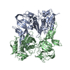
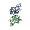
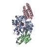
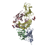
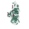
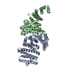
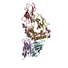
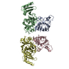
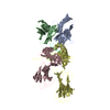
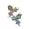
| Title | Crystal structure of the intein homing endonuclease PI-SceI bound to its substrate DNA (Ca2+ free) | ||||||
 Components Components |
| ||||||
 Keywords Keywords | HYDROLASE/DNA / HOMING ENDONUCLEASE / PROTEIN-DNA COMPLEX / INTEIN / ENDONUCLEASE / HYDROLASE-DNA COMPLEX | ||||||
| Function / homology |  Function and homology information Function and homology informationInsulin receptor recycling / Transferrin endocytosis and recycling / ROS and RNS production in phagocytes / Golgi lumen acidification / Amino acids regulate mTORC1 / vacuolar proton-transporting V-type ATPase, V1 domain / endosomal lumen acidification / proton-transporting V-type ATPase complex / intron homing / vacuolar proton-transporting V-type ATPase complex ...Insulin receptor recycling / Transferrin endocytosis and recycling / ROS and RNS production in phagocytes / Golgi lumen acidification / Amino acids regulate mTORC1 / vacuolar proton-transporting V-type ATPase, V1 domain / endosomal lumen acidification / proton-transporting V-type ATPase complex / intron homing / vacuolar proton-transporting V-type ATPase complex / intein-mediated protein splicing / vacuolar acidification / fungal-type vacuole membrane / proton-transporting ATPase activity, rotational mechanism / H+-transporting two-sector ATPase / ATP metabolic process / proton transmembrane transport / endonuclease activity / Hydrolases; Acting on ester bonds / Golgi membrane / hydrolase activity / mRNA binding / DNA binding / ATP binding Similarity search - Function | ||||||
| Biological species |  | ||||||
| Method |  X-RAY DIFFRACTION / SYNCHROTRON / MOLECULAR REPLACEMENT / Resolution: 3.2 Å X-RAY DIFFRACTION / SYNCHROTRON / MOLECULAR REPLACEMENT / Resolution: 3.2 Å | ||||||
 Authors Authors | Moure, C.M. / Gimble, F.S. / Quiocho, F.A. | ||||||
 Citation Citation | Journal: Nat.Struct.Biol. / Year: 2002 Title: Crystal structure of the intein homing endonuclease PI-SceI bound to its recognition sequence. Authors: Moure, C.M. / Gimble, F.S. / Quiocho, F.A. #1: Journal: Cell(Cambridge,Mass.) / Year: 1997Title: Crystal structure of PI-SceI, a homing endonuclease with protein splicing activity Authors: Duan, X. / Gimble, F.S. / Quiocho, F.A. | ||||||
| History |
|
- Structure visualization
| Structure viewer | Molecule: MolmilJmol/JSmol |
|---|
- Downloads & links
-Download
| PDBx/mmCIF format | 1lwt.cif.gz | 142.5 KB | Display | PDBx/mmCIF format |
|---|---|---|---|---|
| PDB format | pdb1lwt.ent.gz | 107.1 KB | Display | PDB format |
| PDBx/mmJSON format | 1lwt.json.gz | Tree view | PDBx/mmJSON format | |
| Others |  Other downloads Other downloads |
-Validation report
| Arichive directory | https://data.pdbj.org/pub/pdb/validation_reports/lw/1lwtftp://data.pdbj.org/pub/pdb/validation_reports/lw/1lwt | HTTPS FTP |
|---|
-Related structure data
| Related structure data |  1lwsSC S: Starting model for refinement C: citing same article ( |
|---|---|
| Similar structure data |

-Links
 PDBj
PDBj


- Assembly
| Deposited unit | 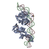
| ||||||||
|---|---|---|---|---|---|---|---|---|---|
| 1 |
| ||||||||
| Unit cell |
|
-Components
| #1: DNA chain | Mass: 11599.467 Da / Num. of mol.: 1 / Source method: obtained synthetically |
|---|---|
| #2: DNA chain | Mass: 11172.185 Da / Num. of mol.: 1 / Source method: obtained synthetically |
| #3: Protein | Mass: 51379.051 Da / Num. of mol.: 1 Source method: isolated from a genetically manipulated source Source: (gene. exp.) Gene: VMA1 / Plasmid: PT7PI-SCEI / Production host:  References: UniProt: P17255, Hydrolases; Acting on ester bonds |
| #4: Water | ChemComp-HOH /  Mass: 18.015 Da / Num. of mol.: 15 / Source method: isolated from a natural source / Formula: H2O Mass: 18.015 Da / Num. of mol.: 15 / Source method: isolated from a natural source / Formula: H2O |
| Has protein modification | Y |
-Experimental details
-Experiment
| Experiment | Method: X-RAY DIFFRACTION / Number of used crystals: 1 |
|---|
- Sample preparation
| Crystal | Density Matthews: 2.83 Å3/Da / Density % sol: 56.49 % | ||||||||||||||||||||||||||||||||||||
|---|---|---|---|---|---|---|---|---|---|---|---|---|---|---|---|---|---|---|---|---|---|---|---|---|---|---|---|---|---|---|---|---|---|---|---|---|---|
| Crystal grow | Temperature: 291 K / Method: vapor diffusion, hanging drop / pH: 5.8 Details: PEG 3550, na citrate, ammonium acetate, pH 5.8, VAPOR DIFFUSION, HANGING DROP, temperature 291K | ||||||||||||||||||||||||||||||||||||
| Components of the solutions |
| ||||||||||||||||||||||||||||||||||||
| Crystal grow | *PLUS Temperature: 18 ℃ | ||||||||||||||||||||||||||||||||||||
| Components of the solutions | *PLUS
|
-Data collection
| Diffraction | Mean temperature: 200 K |
|---|---|
| Diffraction source | Source: SYNCHROTRON / Site: APS  / Beamline: 14-BM-C / Wavelength: 1 Å / Beamline: 14-BM-C / Wavelength: 1 Å |
| Detector | Type: ADSC QUANTUM 4 / Detector: CCD / Date: Nov 27, 2000 |
| Radiation | Protocol: SINGLE WAVELENGTH / Monochromatic (M) / Laue (L): M / Scattering type: x-ray |
| Radiation wavelength | Wavelength: 1 Å / Relative weight: 1 |
| Reflection | Resolution: 3.2→30 Å / Num. all: 12363 / Num. obs: 12363 / % possible obs: 83.6 % / Observed criterion σ(I): -3 / Redundancy: 3.9 % / Rmerge(I) obs: 0.082 / Rsym value: 0.082 / Net I/σ(I): 11.9 |
| Reflection shell | Resolution: 3.2→3.3 Å / Rmerge(I) obs: 0.193 / Num. unique all: 999 / Rsym value: 0.193 / % possible all: 69.8 |
| Reflection | *PLUS Lowest resolution: 25 Å / Num. measured all: 117632 / Rmerge(I) obs: 0.082 |
| Reflection shell | *PLUS % possible obs: 69.9 % / Rmerge(I) obs: 0.193 |
- Processing
| Software |
| ||||||||||||||||||||||||||||||||||||
|---|---|---|---|---|---|---|---|---|---|---|---|---|---|---|---|---|---|---|---|---|---|---|---|---|---|---|---|---|---|---|---|---|---|---|---|---|---|
| Refinement | Method to determine structure: MOLECULAR REPLACEMENT Starting model: PDB ENTRY 1LWS Resolution: 3.2→24.49 Å / Rfactor Rfree error: 0.012 / Isotropic thermal model: isotropic / Cross valid method: THROUGHOUT / σ(F): 2 / Details: BULK SOLVENT MODEL USED
| ||||||||||||||||||||||||||||||||||||
| Solvent computation | Solvent model: FLAT MODEL / Bsol: 10 Å2 / ksol: 0.18428 e/Å3 | ||||||||||||||||||||||||||||||||||||
| Displacement parameters | Biso mean: 36.7 Å2
| ||||||||||||||||||||||||||||||||||||
| Refine analyze | Luzzati coordinate error free: 0.53 Å / Luzzati sigma a free: 0.59 Å | ||||||||||||||||||||||||||||||||||||
| Refinement step | Cycle: LAST / Resolution: 3.2→24.49 Å
| ||||||||||||||||||||||||||||||||||||
| Refine LS restraints |
| ||||||||||||||||||||||||||||||||||||
| LS refinement shell | Resolution: 3.2→3.35 Å / Rfactor Rfree error: 0.4 / Total num. of bins used: 6
| ||||||||||||||||||||||||||||||||||||
| Xplor file |
| ||||||||||||||||||||||||||||||||||||
| Refinement | *PLUS Highest resolution: 3.2 Å / Lowest resolution: 25 Å / % reflection Rfree: 5 % / Rfactor all: 0.248 / Rfactor Rfree: 0.287 / Rfactor Rwork: 0.242 | ||||||||||||||||||||||||||||||||||||
| Solvent computation | *PLUS | ||||||||||||||||||||||||||||||||||||
| Displacement parameters | *PLUS | ||||||||||||||||||||||||||||||||||||
| Refine LS restraints | *PLUS
| ||||||||||||||||||||||||||||||||||||
| LS refinement shell | *PLUS Rfactor Rfree: 0.353 / Rfactor Rwork: 0.301 |
