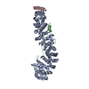
Entry Database : PDB / ID : 1lujTitle Crystal Structure of the Beta-catenin/ICAT Complex Beta-catenin-interacting protein 1 Catenin beta-1 Keywords / / / / Function / homology Function Domain/homology Component
/ / / / / / / / / / / / / / / / / / / / / / / / / / / / / / / / / / / / / / / / / / / / / / / / / / / / / / / / / / / / / / / / / / / / / / / / / / / / / / / / / / / / / / / / / / / / / / / / / / / / / / / / / / / / / / / / / / / / / / / / / / / / / / / / / / / / / Biological species Homo sapiens (human)Mus musculus (house mouse)Method / / / Resolution : 2.5 Å Authors Graham, T.A. / Clements, W.K. / Kimelman, D. / Xu, W. Journal : Mol.Cell / Year : 2002Title : The crystal structure of the beta-catenin/ICAT complex reveals the inhibitory mechanism of ICAT.Authors : Graham, T.A. / Clements, W.K. / Kimelman, D. / Xu, W. History Deposition May 22, 2002 Deposition site / Processing site Revision 1.0 Oct 16, 2002 Provider / Type Revision 1.1 Apr 28, 2008 Group Revision 1.2 Jul 13, 2011 Group Revision 1.3 Mar 16, 2016 Group Revision 1.4 Feb 14, 2024 Group / Database referencesCategory chem_comp_atom / chem_comp_bond ... chem_comp_atom / chem_comp_bond / database_2 / struct_ref_seq_dif Item / _database_2.pdbx_database_accession / _struct_ref_seq_dif.details
Show all Show less
 Movie
Movie Controller


 Open data
 Basic information
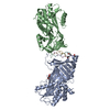
 Components
 Keywords
 Function and homology information
 Homo sapiens (human)
 X-RAY DIFFRACTION /
  Authors
 Citation
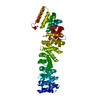
 Structure visualization
 Downloads & links
 Other downloads


 PDBj
PDBj


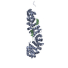
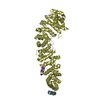
 Assembly

 Sample preparation
 / Beamline: 19-ID / Wavelength: 0.9196 Å
 Processing