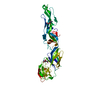
Entry Database : PDB / ID : 1jdhTitle CRYSTAL STRUCTURE OF BETA-CATENIN AND HTCF-4 Keywords / / / Function / homology Function Domain/homology Component
/ / / / / / / / / / / / / / / / / / / / / / / / / / / / / / / / / / / / / / / / / / / / / / / / / / / / / / / / / / / / / / / / / / / / / / / / / / / / / / / / / / / / / / / / / / / / / / / / / / / / / / / / / / / / / / / / / / / / / / / / / / / / / / / / / / / / / / / / / Biological species Homo sapiens (human)Method / / / Resolution : 1.9 Å Authors Graham, T.A. / Ferkey, D.M. / Mao, F. / Kimelman, D. / Xu, W. Journal : Nat.Struct.Biol. / Year : 2001Title : Tcf4 can specifically recognize beta-catenin using alternative conformations.Authors : Graham, T.A. / Ferkey, D.M. / Mao, F. / Kimelman, D. / Xu, W. History Deposition Jun 13, 2001 Deposition site / Processing site Revision 1.0 Dec 5, 2001 Provider / Type Revision 1.1 Apr 27, 2008 Group Revision 1.2 Jul 13, 2011 Group Revision 1.3 Apr 4, 2018 Group / Category / Item Revision 1.4 Feb 7, 2024 Group / Database references / Category / chem_comp_bond / database_2Item / _database_2.pdbx_database_accession
Show all Show less
 Movie
Movie Controller


 Open data
 Basic information
 Components
 Keywords
 Function and homology information
 Homo sapiens (human)
 X-RAY DIFFRACTION /
  Authors
 Citation
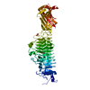
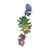
 Structure visualization
 Downloads & links
 Other downloads
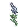


 PDBj
PDBj


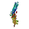
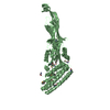
 Assembly
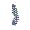

 Mass: 18.015 Da / Num. of mol.: 393 / Source method: isolated from a natural source / Formula: H2O
 Sample preparation
 / Type:
  Processing