 Movie
Movie Controller

[English] 日本語
 Yorodumi
- PDB-1ltz: CRYSTAL STRUCTURE OF CHROMOBACTERIUM VIOLACEUM PHENYLALANINE HYDR... -
+ Open data

- Basic information
| Entry | Database: PDB / ID: 1ltz | ||||||
|---|---|---|---|---|---|---|---|
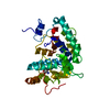
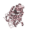
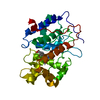
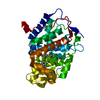
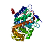
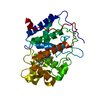
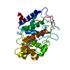
| Title | CRYSTAL STRUCTURE OF CHROMOBACTERIUM VIOLACEUM PHENYLALANINE HYDROXYLASE, STRUCTURE HAS BOUND IRON (III) AND OXIDIZED COFACTOR 7,8-DIHYDROBIOPTERIN | ||||||
 Components Components | PHENYLALANINE-4-HYDROXYLASE | ||||||
 Keywords Keywords | OXIDOREDUCTASE / HYDROXYLASE / PHENYLALANINE / DIHYDROBIOPTERIN / BACTERIAL ENZYME | ||||||
| Function / homology |  Function and homology information Function and homology informationphenylalanine 4-monooxygenase / phenylalanine 4-monooxygenase activity / L-phenylalanine catabolic process / iron ion binding Similarity search - Function | ||||||
| Biological species |  Chromobacterium violaceum (bacteria) Chromobacterium violaceum (bacteria) | ||||||
| Method |  X-RAY DIFFRACTION / SYNCHROTRON / Difference map / Resolution: 1.4 Å X-RAY DIFFRACTION / SYNCHROTRON / Difference map / Resolution: 1.4 Å | ||||||
 Authors Authors | Erlandsen, H. / Kim, J.Y. / Patch, M.G. / Han, A. / Volner, A. / Abu-Omar, M.M. / Stevens, R.C. | ||||||
 Citation Citation | Journal: J.Mol.Biol. / Year: 2002 Title: Structural comparison of bacterial and human iron-dependent phenylalanine hydroxylases: similar fold, different stability and reaction rates. Authors: Erlandsen, H. / Kim, J.Y. / Patch, M.G. / Han, A. / Volner, A. / Abu-Omar, M.M. / Stevens, R.C. | ||||||
| History |
|
- Structure visualization
| Structure viewer | Molecule: MolmilJmol/JSmol |
|---|
- Downloads & links
-Download
| PDBx/mmCIF format | 1ltz.cif.gz | 77.1 KB | Display | PDBx/mmCIF format |
|---|---|---|---|---|
| PDB format | pdb1ltz.ent.gz | 56.1 KB | Display | PDB format |
| PDBx/mmJSON format | 1ltz.json.gz | Tree view | PDBx/mmJSON format | |
| Others |  Other downloads Other downloads |
-Validation report
| Arichive directory | https://data.pdbj.org/pub/pdb/validation_reports/lt/1ltzftp://data.pdbj.org/pub/pdb/validation_reports/lt/1ltz | HTTPS FTP |
|---|
-Related structure data
| Related structure data |  1ltuSC  1ltvC S: Starting model for refinement C: citing same article ( |
|---|---|
| Similar structure data |


-Links
 PDBj
PDBj


- Assembly
| Deposited unit | 
| ||||||||
|---|---|---|---|---|---|---|---|---|---|
| 1 |
| ||||||||
| Unit cell |
|
-Components
| #1: Protein | Mass: 33627.969 Da / Num. of mol.: 1 Source method: isolated from a genetically manipulated source Source: (gene. exp.) Chromobacterium violaceum (bacteria) / Plasmid: pET-3a / Production host: |
|---|---|
| #2: Chemical | ChemComp-FE /   Mass: 55.845 Da / Num. of mol.: 1 / Source method: obtained synthetically / Formula: Fe Mass: 55.845 Da / Num. of mol.: 1 / Source method: obtained synthetically / Formula: Fe |
| #3: Chemical | ChemComp-CL /   Mass: 35.453 Da / Num. of mol.: 1 / Source method: obtained synthetically / Formula: Cl Mass: 35.453 Da / Num. of mol.: 1 / Source method: obtained synthetically / Formula: Cl |
| #4: Chemical | ChemComp-HBI /   Mass: 239.231 Da / Num. of mol.: 1 / Source method: obtained synthetically / Formula: C9H13N5O3 Mass: 239.231 Da / Num. of mol.: 1 / Source method: obtained synthetically / Formula: C9H13N5O3 |
| #5: Water | ChemComp-HOH /  Mass: 18.015 Da / Num. of mol.: 344 / Source method: isolated from a natural source / Formula: H2O Mass: 18.015 Da / Num. of mol.: 344 / Source method: isolated from a natural source / Formula: H2O |
-Experimental details
-Experiment
| Experiment | Method: X-RAY DIFFRACTION / Number of used crystals: 1 |
|---|
- Sample preparation
| Crystal | Density Matthews: 2.15 Å3/Da / Density % sol: 42.89 % | |||||||||||||||||||||||||||||||||||
|---|---|---|---|---|---|---|---|---|---|---|---|---|---|---|---|---|---|---|---|---|---|---|---|---|---|---|---|---|---|---|---|---|---|---|---|---|
| Crystal grow | Temperature: 277.15 K / Method: vapor diffusion, hanging drop / pH: 7.5 Details: ammonium sulfate, NaCl, HEPES buffer, pH 7.5, VAPOR DIFFUSION, HANGING DROP, temperature 277.15K | |||||||||||||||||||||||||||||||||||
| Crystal grow | *PLUS Temperature: 4 ℃ | |||||||||||||||||||||||||||||||||||
| Components of the solutions | *PLUS
|
-Data collection
| Diffraction | Mean temperature: 100 K |
|---|---|
| Diffraction source | Source: SYNCHROTRON / Site: ALS  / Beamline: 5.0.2 / Wavelength: 1 Å / Beamline: 5.0.2 / Wavelength: 1 Å |
| Detector | Type: ADSC QUANTUM 4 / Detector: CCD / Date: Nov 8, 2000 |
| Radiation | Protocol: SINGLE WAVELENGTH / Monochromatic (M) / Laue (L): M / Scattering type: x-ray |
| Radiation wavelength | Wavelength: 1 Å / Relative weight: 1 |
| Reflection | Resolution: 1.4→20 Å / Num. all: 54370 / Num. obs: 54730 / % possible obs: 95.2 % / Observed criterion σ(F): 0 / Observed criterion σ(I): 0 / Redundancy: 9 % / Rmerge(I) obs: 0.057 / Net I/σ(I): 28 |
| Reflection shell | Resolution: 1.4→1.42 Å / Redundancy: 3 % / Rmerge(I) obs: 0.427 / Mean I/σ(I) obs: 2.4 / Num. unique all: 1910 / % possible all: 67.2 |
| Reflection | *PLUS Highest resolution: 1.4 Å / Lowest resolution: 20 Å / Redundancy: 9-12 / Num. measured all: 54730 / Rmerge(I) obs: 0.057 |
| Reflection shell | *PLUS % possible obs: 67.2 % / Rmerge(I) obs: 0.427 |
- Processing
| Software |
| |||||||||||||||||||||||||
|---|---|---|---|---|---|---|---|---|---|---|---|---|---|---|---|---|---|---|---|---|---|---|---|---|---|---|
| Refinement | Method to determine structure: Difference map Starting model: PDB ENTRY 1LTU Resolution: 1.4→20 Å / Isotropic thermal model: anisotropic / Cross valid method: THROUGHOUT / σ(F): 0 / σ(I): 0 / Stereochemistry target values: Engh & Huber
| |||||||||||||||||||||||||
| Refine analyze |
| |||||||||||||||||||||||||
| Refinement step | Cycle: LAST / Resolution: 1.4→20 Å
| |||||||||||||||||||||||||
| Refine LS restraints |
| |||||||||||||||||||||||||
| Software | *PLUS Name: 'CNS, SHELZ' / Version: 97 / Classification: refinement | |||||||||||||||||||||||||
| Refinement | *PLUS Highest resolution: 1.4 Å / Lowest resolution: 20 Å / Rfactor Rfree: 0.223 / Rfactor Rwork: 0.159 | |||||||||||||||||||||||||
| Solvent computation | *PLUS | |||||||||||||||||||||||||
| Displacement parameters | *PLUS | |||||||||||||||||||||||||
| Refine LS restraints | *PLUS
| |||||||||||||||||||||||||
| LS refinement shell | *PLUS Highest resolution: 1.4 Å / Lowest resolution: 1.42 Å |
