 Movie
Movie Controller

[English] 日本語
 Yorodumi
- PDB-1lld: MOLECULAR BASIS OF ALLOSTERIC ACTIVATION OF BACTERIAL L-LACTATE D... -
+ Open data

- Basic information
| Entry | Database: PDB / ID: 1lld | ||||||
|---|---|---|---|---|---|---|---|
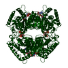
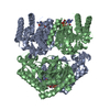
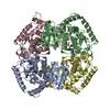
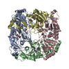
| Title | MOLECULAR BASIS OF ALLOSTERIC ACTIVATION OF BACTERIAL L-LACTATE DEHYDROGENASE | ||||||
 Components Components | L-LACTATE DEHYDROGENASE | ||||||
 Keywords Keywords | OXIDOREDUCTASE / OXIDOREDUCTASE(CHOH (D)-NAD (A)) | ||||||
| Function / homology |  Function and homology information Function and homology informationL-lactate dehydrogenase / L-lactate dehydrogenase (NAD+) activity / lactate metabolic process / glycolytic process / cytoplasm Similarity search - Function | ||||||
| Biological species |  Bifidobacterium longum subsp. longum (bacteria) Bifidobacterium longum subsp. longum (bacteria) | ||||||
| Method |  X-RAY DIFFRACTION / Resolution: 2 Å X-RAY DIFFRACTION / Resolution: 2 Å | ||||||
 Authors Authors | Iwata, S. / Ohta, T. | ||||||
 Citation Citation | Journal: J.Mol.Biol. / Year: 1993 Title: Molecular basis of allosteric activation of bacterial L-lactate dehydrogenase. Authors: Iwata, S. / Ohta, T. #1: Journal: FARADAY DISC.CHEM.SOC / Year: 1992Title: Mechanism of Allosteric Transition of Bacterial L-Lactate Dehydrogenase Authors: Iwata, S. / Ohta, T. #2: Journal: Agric.Biol.Chem. / Year: 1989Title: Amino Acid Residues in the Allosteric Site of L-Lactate Dehydrogenase from Bifidobacterium Longum Authors: Iwata, S. / Minowa, T. / Sakai, H. / Ohta, T. #3: Journal: Gene / Year: 1989Title: Sequence and Characteristics of the Bifidobacterium Longum Gene Encoding L-Lactate Dehydrogenase and the Primary Structure of the Enzyme: A New Feature of the Allosteric Site Authors: Minowa, T. / Iwata, S. / Sakai, H. / Masaki, H. / Ohta, T. #4: Journal: J.Biochem.(Tokyo) / Year: 1989Title: Crystallization of and Preliminary Crystallographic Data for Allosteric L-Lactate Dehydrogenase from Bifidobacterium Longum Authors: Wata, S. / Minowa, T. / Mikami, B. / Morita, Y. / Ohta, T. | ||||||
| History |
| ||||||
| Remark 650 | HELIX THE HELIX NAMES USED IN EARLIER LDH ENTRIES BY M.G.ROSSMANN ET AL. ARE GIVEN IN THE REMARK ...HELIX THE HELIX NAMES USED IN EARLIER LDH ENTRIES BY M.G.ROSSMANN ET AL. ARE GIVEN IN THE REMARK FIELD OF THE HELIX RECORDS. |
- Structure visualization
| Structure viewer | Molecule: MolmilJmol/JSmol |
|---|
- Downloads & links
-Download
| PDBx/mmCIF format | 1lld.cif.gz | 133.7 KB | Display | PDBx/mmCIF format |
|---|---|---|---|---|
| PDB format | pdb1lld.ent.gz | 106.3 KB | Display | PDB format |
| PDBx/mmJSON format | 1lld.json.gz | Tree view | PDBx/mmJSON format | |
| Others |  Other downloads Other downloads |
-Validation report
| Arichive directory | https://data.pdbj.org/pub/pdb/validation_reports/ll/1lldftp://data.pdbj.org/pub/pdb/validation_reports/ll/1lld | HTTPS FTP |
|---|
-Related structure data
| Similar structure data |
|---|
-Links
 PDBj
PDBj


- Assembly
| Deposited unit | 
| ||||||||
|---|---|---|---|---|---|---|---|---|---|
| 1 | 
| ||||||||
| Unit cell |
| ||||||||
| Atom site foot note | 1: CIS PROLINE - PRO A 126 / 2: CIS PROLINE - PRO B 126 3: RESIDUES A 86 - A 88, B 86 - B 93, A 232 - A 234, AND B 232 - B 234 ARE DISORDERED. ALTHOUGH THE COORDINATES OF THESE RESIDUES ARE INCLUDED IN THIS ENTRY, THEY ARE LESS RELIABLE THAN THOSE OF OTHER RESIDUES. | ||||||||
| Noncrystallographic symmetry (NCS) | NCS oper: (Code: given Matrix: (-0.309, 0.951, -0.003), Vector: Details | THE CRYSTALLOGRAPHIC ASYMMETRIC UNIT CONTAINS TWO SUBUNITS OF THE TETRAMER. THESE SUBUNITS HAVE BEEN ASSIGNED CHAIN IDENTIFIERS "A" AND "B". THE RESIDUES IN EACH CHAIN ARE NUMBERED SEQUENTIALLY FROM 1 - 319. THERE IS NO SIGNIFICANT DIFFERENCE BETWEEN THE TWO SUBUNIT STRUCTURES. THE TETRAMER CAN BE GENERATED FROM THE DIMER IN THIS ENTRY BY ROTATING 180 DEGREES ABOUT THE CRYSTALLOGRAPHIC DIAD PARALLEL TO Z AXIS THROUGH THE POINT (1/2,1/2,1/2). THE TRANSFORMATION PRESENTED ON *MTRIX* RECORDS BELOW WILL YIELD APPROXIMATE COORDINATES FOR CHAIN *B* WHEN APPLIED TO CHAIN *A*. | |
-Components
| #1: Protein | Mass: 34128.777 Da / Num. of mol.: 2 Source method: isolated from a genetically manipulated source Source: (gene. exp.) Bifidobacterium longum subsp. longum (bacteria)Species: Bifidobacterium longum / Strain: subsp. longum References: UniProt: P19869, UniProt: E8ME30*PLUS, L-lactate dehydrogenase #2: Chemical |   Mass: 663.425 Da / Num. of mol.: 2 / Source method: obtained synthetically / Formula: C21H27N7O14P2 / Comment: NAD*YM Mass: 663.425 Da / Num. of mol.: 2 / Source method: obtained synthetically / Formula: C21H27N7O14P2 / Comment: NAD*YM#3: Water | ChemComp-HOH / |  Mass: 18.015 Da / Num. of mol.: 222 / Source method: isolated from a natural source / Formula: H2O Mass: 18.015 Da / Num. of mol.: 222 / Source method: isolated from a natural source / Formula: H2OCompound details | THIS ENZYME IS A MUTANT WITH CYS 199 REPLACED BY SER. THIS MUTATION IS FOR THE HEAVY-ATOM ...THIS ENZYME IS A MUTANT WITH CYS 199 REPLACED BY SER. THIS MUTATION IS FOR THE HEAVY-ATOM DERIVATIVE | |
|---|
-Experimental details
-Experiment
| Experiment | Method: X-RAY DIFFRACTION |
|---|
- Sample preparation
| Crystal | Density Matthews: 3.25 Å3/Da / Density % sol: 62.12 % | ||||||||||||||||||||||||||||||||||||||||||||||||
|---|---|---|---|---|---|---|---|---|---|---|---|---|---|---|---|---|---|---|---|---|---|---|---|---|---|---|---|---|---|---|---|---|---|---|---|---|---|---|---|---|---|---|---|---|---|---|---|---|---|
| Crystal grow | *PLUS pH: 7 / Method: vapor diffusion, hanging dropDetails: taken from Iwata, S. et al (1989). J. Biochem., 106-558-559. | ||||||||||||||||||||||||||||||||||||||||||||||||
| Components of the solutions | *PLUS
|
-Data collection
| Radiation | Scattering type: x-ray |
|---|---|
| Radiation wavelength | Relative weight: 1 |
| Reflection | *PLUS Highest resolution: 1.8 Å / Num. obs: 64557 / % possible obs: 77 % / Rmerge(I) obs: 0.061 |
- Processing
| Software | Name: PROLSQ / Classification: refinement | ||||||||||||||||||||||||||||||||||||||||||||||||||||||||||||||||||||||||||||||||||||
|---|---|---|---|---|---|---|---|---|---|---|---|---|---|---|---|---|---|---|---|---|---|---|---|---|---|---|---|---|---|---|---|---|---|---|---|---|---|---|---|---|---|---|---|---|---|---|---|---|---|---|---|---|---|---|---|---|---|---|---|---|---|---|---|---|---|---|---|---|---|---|---|---|---|---|---|---|---|---|---|---|---|---|---|---|---|
| Refinement | Resolution: 2→10 Å / σ(F): 1 Details: RESIDUES A 86 - A 88, B 86 - B 93, A 232 - A 234, AND B 232 - B 234 ARE DISORDERED. ALTHOUGH THE COORDINATES OF THESE RESIDUES ARE INCLUDED IN THIS ENTRY, THEY ARE LESS RELIABLE THAN THOSE OF OTHER RESIDUES.
| ||||||||||||||||||||||||||||||||||||||||||||||||||||||||||||||||||||||||||||||||||||
| Refinement step | Cycle: LAST / Resolution: 2→10 Å
| ||||||||||||||||||||||||||||||||||||||||||||||||||||||||||||||||||||||||||||||||||||
| Refine LS restraints |
| ||||||||||||||||||||||||||||||||||||||||||||||||||||||||||||||||||||||||||||||||||||
| Software | *PLUS Name: PROLSQ / Classification: refinement | ||||||||||||||||||||||||||||||||||||||||||||||||||||||||||||||||||||||||||||||||||||
| Refinement | *PLUS Highest resolution: 2 Å / Lowest resolution: 10 Å / Num. reflection obs: 47766 / σ(F): 1 / Rfactor obs: 0.179 | ||||||||||||||||||||||||||||||||||||||||||||||||||||||||||||||||||||||||||||||||||||
| Solvent computation | *PLUS | ||||||||||||||||||||||||||||||||||||||||||||||||||||||||||||||||||||||||||||||||||||
| Displacement parameters | *PLUS |
