 Movie
Movie Controller

[English] 日本語
 Yorodumi
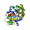
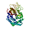
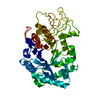
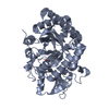
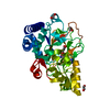
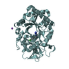
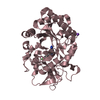
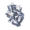
- PDB-1lg2: CRYSTAL STRUCTURE OF HUMAN CHITOTRIOSIDASE IN COMPLEX WITH ETHYLE... -
+ Open data

- Basic information
| Entry | Database: PDB / ID: 1lg2 | ||||||
|---|---|---|---|---|---|---|---|
| Title | CRYSTAL STRUCTURE OF HUMAN CHITOTRIOSIDASE IN COMPLEX WITH ETHYLENE GLYCOL | ||||||
 Components Components | chitotriosidase | ||||||
 Keywords Keywords | HYDROLASE / CHITINASE / CHITIN / GAUCHER | ||||||
| Function / homology |  Function and homology information Function and homology informationpolysaccharide digestion / Digestion of dietary carbohydrate / chitinase activity / endochitinase activity / chitinase / chitin catabolic process / chitin binding / polysaccharide catabolic process / hydrolase activity, hydrolyzing O-glycosyl compounds / response to bacterium ...polysaccharide digestion / Digestion of dietary carbohydrate / chitinase activity / endochitinase activity / chitinase / chitin catabolic process / chitin binding / polysaccharide catabolic process / hydrolase activity, hydrolyzing O-glycosyl compounds / response to bacterium / specific granule lumen / tertiary granule lumen / lysosome / immune response / Neutrophil degranulation / : / extracellular region Similarity search - Function | ||||||
| Biological species |  Homo sapiens (human) Homo sapiens (human) | ||||||
| Method |  X-RAY DIFFRACTION / MOLECULAR REPLACEMENT / Resolution: 2.1 Å X-RAY DIFFRACTION / MOLECULAR REPLACEMENT / Resolution: 2.1 Å | ||||||
 Authors Authors | Fusetti, F. / Rozeboom, H.J. / Dijkstra, B.W. | ||||||
 Citation Citation | Journal: J.Biol.Chem. / Year: 2002 Title: Structure of Human Chitotriosidase. Implications for Specific Inhibitor Design and Function of Mammalian Chitinase-Like Lectins. Authors: Fusetti, F. / Von Moeller, H. / Houston, D. / Rozeboom, H.J. / Dijkstra, B.W. / Boot, R.G. / Aerts, J.M. / Van Aalten, D.M. | ||||||
| History |
|
- Structure visualization
| Structure viewer | Molecule: MolmilJmol/JSmol |
|---|
- Downloads & links
-Download
| PDBx/mmCIF format | 1lg2.cif.gz | 88.6 KB | Display | PDBx/mmCIF format |
|---|---|---|---|---|
| PDB format | pdb1lg2.ent.gz | 64.4 KB | Display | PDB format |
| PDBx/mmJSON format | 1lg2.json.gz | Tree view | PDBx/mmJSON format | |
| Others |  Other downloads Other downloads |
-Validation report
| Arichive directory | https://data.pdbj.org/pub/pdb/validation_reports/lg/1lg2ftp://data.pdbj.org/pub/pdb/validation_reports/lg/1lg2 | HTTPS FTP |
|---|
-Related structure data


-Links
 PDBj
PDBj

- Assembly
| Deposited unit | 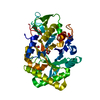
| ||||||||
|---|---|---|---|---|---|---|---|---|---|
| 1 |
| ||||||||
| Unit cell |
|
-Components
| #1: Protein | Mass: 40784.695 Da / Num. of mol.: 1 / Fragment: residues 22-386 Source method: isolated from a genetically manipulated source Source: (gene. exp.) Homo sapiens (human) / Cell line (production host): BHK / Production host:  Mesocricetus auratus (golden hamster) / References: UniProt: Q13231 Mesocricetus auratus (golden hamster) / References: UniProt: Q13231 | ||||
|---|---|---|---|---|---|
| #2: Chemical | ChemComp-EDO /   Mass: 62.068 Da / Num. of mol.: 5 / Source method: obtained synthetically / Formula: C2H6O2 Mass: 62.068 Da / Num. of mol.: 5 / Source method: obtained synthetically / Formula: C2H6O2#3: Water | ChemComp-HOH / |  Mass: 18.015 Da / Num. of mol.: 201 / Source method: isolated from a natural source / Formula: H2O Mass: 18.015 Da / Num. of mol.: 201 / Source method: isolated from a natural source / Formula: H2OHas protein modification | Y | |
-Experimental details
-Experiment
| Experiment | Method: X-RAY DIFFRACTION / Number of used crystals: 1 |
|---|
- Sample preparation
| Crystal | Density Matthews: 2.26 Å3/Da / Density % sol: 45.2 % | ||||||||||||||||||||||||||||||
|---|---|---|---|---|---|---|---|---|---|---|---|---|---|---|---|---|---|---|---|---|---|---|---|---|---|---|---|---|---|---|---|
| Crystal grow | pH: 5.6 / Details: pH 5.6 | ||||||||||||||||||||||||||||||
| Crystal grow | *PLUS pH: 8.7 / Method: vapor diffusion | ||||||||||||||||||||||||||||||
| Components of the solutions | *PLUS
|
-Data collection
| Diffraction | Mean temperature: 100 K |
|---|---|
| Diffraction source | Source: ROTATING ANODE / Wavelength: 1.5418 |
| Detector | Type: MAC Science DIP-2030 / Detector: IMAGE PLATE / Date: Jul 1, 2001 |
| Radiation | Monochromator: MIRRORS / Protocol: SINGLE WAVELENGTH / Monochromatic (M) / Laue (L): M / Scattering type: x-ray |
| Radiation wavelength | Wavelength: 1.5418 Å / Relative weight: 1 |
| Reflection | Resolution: 2.1→50 Å / Num. obs: 20542 / % possible obs: 99.6 % / Observed criterion σ(I): -3 / Redundancy: 3.5 % / Biso Wilson estimate: 22.184 Å2 / Rmerge(I) obs: 0.072 / Net I/σ(I): 18.9 |
| Reflection shell | Resolution: 2.1→2.15 Å / Redundancy: 3.3 % / Rmerge(I) obs: 0.225 / Mean I/σ(I) obs: 5.9 / Num. unique all: 1330 / % possible all: 97 |
| Reflection | *PLUS Highest resolution: 2.1 Å / Lowest resolution: 13.4 Å / Num. obs: 20533 / Redundancy: 3.6 % / Num. measured all: 73055 |
| Reflection shell | *PLUS % possible obs: 97 % / Redundancy: 3.2 % / Num. unique obs: 1330 / Num. measured obs: 4290 / Mean I/σ(I) obs: 4.6 |
- Processing
| Software |
| ||||||||||||||||||||||||||||||||||||||||||||||||||||||||||||||||||||||||||||||||
|---|---|---|---|---|---|---|---|---|---|---|---|---|---|---|---|---|---|---|---|---|---|---|---|---|---|---|---|---|---|---|---|---|---|---|---|---|---|---|---|---|---|---|---|---|---|---|---|---|---|---|---|---|---|---|---|---|---|---|---|---|---|---|---|---|---|---|---|---|---|---|---|---|---|---|---|---|---|---|---|---|---|
| Refinement | Method to determine structure: MOLECULAR REPLACEMENT Starting model: HUMAN CHITOTRIOSIADSE TETRAGONAL CRYSTAL FORM Resolution: 2.1→23.64 Å / Rfactor Rfree error: 0.005 / Data cutoff high absF: 1089189.58 / Data cutoff high rms absF: 1089189.58 / Data cutoff low absF: 0 / Isotropic thermal model: RESTRAINED / Cross valid method: THROUGHOUT / σ(F): 0 / Stereochemistry target values: ENGH & HUBER
| ||||||||||||||||||||||||||||||||||||||||||||||||||||||||||||||||||||||||||||||||
| Solvent computation | Solvent model: FLAT MODEL / Bsol: 52.1673 Å2 / ksol: 0.395364 e/Å3 | ||||||||||||||||||||||||||||||||||||||||||||||||||||||||||||||||||||||||||||||||
| Displacement parameters | Biso mean: 19.6 Å2
| ||||||||||||||||||||||||||||||||||||||||||||||||||||||||||||||||||||||||||||||||
| Refine analyze | Luzzati coordinate error free: 0.26 Å / Luzzati sigma a free: 0.13 Å | ||||||||||||||||||||||||||||||||||||||||||||||||||||||||||||||||||||||||||||||||
| Refinement step | Cycle: LAST / Resolution: 2.1→23.64 Å
| ||||||||||||||||||||||||||||||||||||||||||||||||||||||||||||||||||||||||||||||||
| Refine LS restraints |
| ||||||||||||||||||||||||||||||||||||||||||||||||||||||||||||||||||||||||||||||||
| LS refinement shell | Resolution: 2.1→2.23 Å / Rfactor Rfree error: 0.013 / Total num. of bins used: 6
| ||||||||||||||||||||||||||||||||||||||||||||||||||||||||||||||||||||||||||||||||
| Xplor file |
| ||||||||||||||||||||||||||||||||||||||||||||||||||||||||||||||||||||||||||||||||
| Refinement | *PLUS Highest resolution: 2.1 Å / Lowest resolution: 13.4 Å / Rfactor Rfree: 0.227 / Rfactor Rwork: 0.204 | ||||||||||||||||||||||||||||||||||||||||||||||||||||||||||||||||||||||||||||||||
| Solvent computation | *PLUS | ||||||||||||||||||||||||||||||||||||||||||||||||||||||||||||||||||||||||||||||||
| Displacement parameters | *PLUS | ||||||||||||||||||||||||||||||||||||||||||||||||||||||||||||||||||||||||||||||||
| Refine LS restraints | *PLUS
|
