 Movie
Movie Controller

[English] 日本語
 Yorodumi
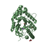
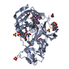
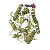
- PDB-3emq: Crystal structure of xilanase XynB from Paenibacillus barcelonens... -
+ Open data

- Basic information
| Entry | Database: PDB / ID: 3emq | ||||||
|---|---|---|---|---|---|---|---|
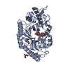
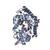
| Title | Crystal structure of xilanase XynB from Paenibacillus barcelonensis complexed with an inhibitor | ||||||
 Components Components | Endo-1,4-beta-xylanase | ||||||
 Keywords Keywords | HYDROLASE / (alpha/beta)8 barrel | ||||||
| Function / homology |  Function and homology information Function and homology informationendo-1,4-beta-xylanase / endo-1,4-beta-xylanase activity / xylan catabolic process / cytoplasm Similarity search - Function | ||||||
| Biological species |  | ||||||
| Method |  X-RAY DIFFRACTION / MOLECULAR REPLACEMENT / molecular replacement / Resolution: 2.73 Å X-RAY DIFFRACTION / MOLECULAR REPLACEMENT / molecular replacement / Resolution: 2.73 Å | ||||||
 Authors Authors | Sanz-Aparicio, J. / Isorna, P. | ||||||
 Citation Citation | Journal: J.Biol.Chem. / Year: 2010 Title: Structural insights into the specificity of Xyn10B from Paenibacillus barcinonensis and its improved stability by forced protein evolution. Authors: Gallardo, O. / Pastor, F.I. / Polaina, J. / Diaz, P. / Lysek, R. / Vogel, P. / Isorna, P. / Gonzalez, B. / Sanz-Aparicio, J. | ||||||
| History |
|
- Structure visualization
| Structure viewer | Molecule: MolmilJmol/JSmol |
|---|
- Downloads & links
-Download
| PDBx/mmCIF format | 3emq.cif.gz | 86.6 KB | Display | PDBx/mmCIF format |
|---|---|---|---|---|
| PDB format | pdb3emq.ent.gz | 63.6 KB | Display | PDB format |
| PDBx/mmJSON format | 3emq.json.gz | Tree view | PDBx/mmJSON format | |
| Others |  Other downloads Other downloads |
-Validation report
| Arichive directory | https://data.pdbj.org/pub/pdb/validation_reports/em/3emqftp://data.pdbj.org/pub/pdb/validation_reports/em/3emq | HTTPS FTP |
|---|
-Related structure data
| Related structure data | 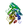 3emcSC 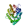 3emzC S: Starting model for refinement C: citing same article ( |
|---|---|
| Similar structure data |


-Links
 PDBj
PDBj
- Assembly
| Deposited unit | 
| ||||||||
|---|---|---|---|---|---|---|---|---|---|
| 1 |
| ||||||||
| Unit cell |
|
-Components
| #1: Protein | Mass: 38475.855 Da / Num. of mol.: 1 Source method: isolated from a genetically manipulated source Source: (gene. exp.) |
|---|---|
| #2: Chemical | ChemComp-HAH / (  Mass: 251.278 Da / Num. of mol.: 1 / Source method: obtained synthetically / Formula: C13H17NO4 Mass: 251.278 Da / Num. of mol.: 1 / Source method: obtained synthetically / Formula: C13H17NO4 |
| #3: Water | ChemComp-HOH /  Mass: 18.015 Da / Num. of mol.: 265 / Source method: isolated from a natural source / Formula: H2O Mass: 18.015 Da / Num. of mol.: 265 / Source method: isolated from a natural source / Formula: H2O |
-Experimental details
-Experiment
| Experiment | Method: X-RAY DIFFRACTION / Number of used crystals: 1 |
|---|
- Sample preparation
| Crystal | Density Matthews: 2.15 Å3/Da / Density % sol: 42.68 % |
|---|---|
| Crystal grow | Temperature: 298 K / Method: vapor diffusion / pH: 7 Details: 35% (v/v) MPD, 17% (w/v) PEG 3350, 100 mM tris, 10mM inhibitor , VAPOR DIFFUSION, temperature 298K |
-Data collection
| Diffraction | Mean temperature: 120 K |
|---|---|
| Diffraction source | Source: ROTATING ANODE / Type: ENRAF-NONIUS FR591 / Wavelength: 1.5418 Å |
| Detector | Type: Nonius Kappa CCD / Detector: CCD / Date: Nov 23, 2007 / Details: osmic mirrors |
| Radiation | Monochromator: osmic mirrors / Protocol: SINGLE WAVELENGTH / Monochromatic (M) / Laue (L): M / Scattering type: x-ray |
| Radiation wavelength | Wavelength: 1.5418 Å / Relative weight: 1 |
| Reflection | Resolution: 2.61→50 Å / Num. all: 10538 / Num. obs: 10038 / % possible obs: 90 % / Observed criterion σ(F): 0 / Observed criterion σ(I): 0 / Redundancy: 5.8 % / Biso Wilson estimate: 38.6 Å2 / Rmerge(I) obs: 0.103 / Net I/σ(I): 10.2 |
| Reflection shell | Resolution: 2.61→2.73 Å / Redundancy: 3.2 % / Rmerge(I) obs: 0.65 / Mean I/σ(I) obs: 2.4 / Num. unique all: 1261 / Rsym value: 0.65 / % possible all: 11 |
-Phasing
| Phasing | Method: molecular replacement |
|---|
- Processing
| Software |
| ||||||||||||||||||||||||||||
|---|---|---|---|---|---|---|---|---|---|---|---|---|---|---|---|---|---|---|---|---|---|---|---|---|---|---|---|---|---|
| Refinement | Method to determine structure: MOLECULAR REPLACEMENT Starting model: 3EMC Resolution: 2.73→16.92 Å / Occupancy max: 1 / Occupancy min: 1 / Isotropic thermal model: Isotropic / Cross valid method: THROUGHOUT / σ(F): 0 / Stereochemistry target values: Engh & Huber
| ||||||||||||||||||||||||||||
| Solvent computation | Bsol: 59.348 Å2 | ||||||||||||||||||||||||||||
| Displacement parameters | Biso max: 66.58 Å2 / Biso mean: 25.734 Å2 / Biso min: 1.04 Å2
| ||||||||||||||||||||||||||||
| Refinement step | Cycle: LAST / Resolution: 2.73→16.92 Å
| ||||||||||||||||||||||||||||
| Refine LS restraints |
| ||||||||||||||||||||||||||||
| LS refinement shell | Resolution: 2.73→2.8 Å /
| ||||||||||||||||||||||||||||
| Xplor file |
|
