 Movie
Movie Controller

[English] 日本語
 Yorodumi
- PDB-1ld3: Crystal Structure of B. subilis ferrochelatase with Zn(2+) bound ... -
+ Open data

- Basic information
| Entry | Database: PDB / ID: 1ld3 | ||||||
|---|---|---|---|---|---|---|---|
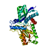
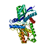
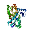
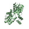
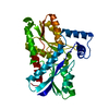
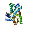
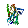
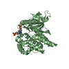
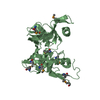
| Title | Crystal Structure of B. subilis ferrochelatase with Zn(2+) bound at the active site. | ||||||
 Components Components | Ferrochelatase | ||||||
 Keywords Keywords | LYASE / PI-HELIX / ROSSMANN FOLD | ||||||
| Function / homology |  Function and homology information Function and homology informationcoproporphyrin ferrochelatase / protoporphyrin ferrochelatase activity / heme biosynthetic process / metal ion binding / cytoplasm Similarity search - Function | ||||||
| Biological species |  | ||||||
| Method |  X-RAY DIFFRACTION / SYNCHROTRON / MOLECULAR REPLACEMENT / Resolution: 2.6 Å X-RAY DIFFRACTION / SYNCHROTRON / MOLECULAR REPLACEMENT / Resolution: 2.6 Å | ||||||
 Authors Authors | Lecerof, D. / Fodje, M.N. / Leon, R.A. / Olsson, U. / Hansson, A. / Sigfridsson, E. / Ryde, U. / Hansson, M. / Al-Karadaghi, S. | ||||||
 Citation Citation | Journal: J.Biol.Inorg.Chem. / Year: 2003 Title: Metal binding to Bacillus subtilis ferrochelatase and interaction between metal sites Authors: Lecerof, D. / Fodje, M.N. / Leon, R.A. / Olsson, U. / Hansson, A. / Sigfridsson, E. / Ryde, U. / Hansson, M. / Al-Karadaghi, S. | ||||||
| History |
|
- Structure visualization
| Structure viewer | Molecule: MolmilJmol/JSmol |
|---|
- Downloads & links
-Download
| PDBx/mmCIF format | 1ld3.cif.gz | 75.1 KB | Display | PDBx/mmCIF format |
|---|---|---|---|---|
| PDB format | pdb1ld3.ent.gz | 54.9 KB | Display | PDB format |
| PDBx/mmJSON format | 1ld3.json.gz | Tree view | PDBx/mmJSON format | |
| Others |  Other downloads Other downloads |
-Validation report
| Arichive directory | https://data.pdbj.org/pub/pdb/validation_reports/ld/1ld3ftp://data.pdbj.org/pub/pdb/validation_reports/ld/1ld3 | HTTPS FTP |
|---|
-Related structure data
| Related structure data |  1n0iC  1dozS S: Starting model for refinement C: citing same article ( |
|---|---|
| Similar structure data |


-Links
 PDBj
PDBj- Assembly
| Deposited unit | 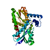
| ||||||||
|---|---|---|---|---|---|---|---|---|---|
| 1 |
| ||||||||
| Unit cell |
|
-Components
| #1: Protein | Mass: 35389.707 Da / Num. of mol.: 1 Source method: isolated from a genetically manipulated source Source: (gene. exp.) |
|---|---|
| #2: Chemical | ChemComp-ZN /   Mass: 65.409 Da / Num. of mol.: 1 / Source method: obtained synthetically / Formula: Zn Mass: 65.409 Da / Num. of mol.: 1 / Source method: obtained synthetically / Formula: Zn |
| #3: Water | ChemComp-HOH /  Mass: 18.015 Da / Num. of mol.: 37 / Source method: isolated from a natural source / Formula: H2O Mass: 18.015 Da / Num. of mol.: 37 / Source method: isolated from a natural source / Formula: H2O |
-Experimental details
-Experiment
| Experiment | Method: X-RAY DIFFRACTION / Number of used crystals: 1 |
|---|
- Sample preparation
| Crystal | Density Matthews: 2.03 Å3/Da / Density % sol: 39.44 % | ||||||||||||||||||||||||||||||||||||||||||||||||
|---|---|---|---|---|---|---|---|---|---|---|---|---|---|---|---|---|---|---|---|---|---|---|---|---|---|---|---|---|---|---|---|---|---|---|---|---|---|---|---|---|---|---|---|---|---|---|---|---|---|
| Crystal grow | Temperature: 298 K / Method: vapor diffusion, hanging drop / pH: 8.5 Details: PEG 2000, magnesium chloride, Tris, pH 8.5, VAPOR DIFFUSION, HANGING DROP, temperature 298K | ||||||||||||||||||||||||||||||||||||||||||||||||
| Crystal grow | *PLUS Details: Hansson, M., (1995) Proteins: Struct.,Funct., Genet., 23, 607. | ||||||||||||||||||||||||||||||||||||||||||||||||
| Components of the solutions | *PLUS
|
-Data collection
| Diffraction | Mean temperature: 100 K |
|---|---|
| Diffraction source | Source: SYNCHROTRON / Site: MAX II  / Beamline: I711 / Wavelength: 0.986 Å / Beamline: I711 / Wavelength: 0.986 Å |
| Detector | Type: MARRESEARCH / Detector: IMAGE PLATE / Date: Oct 25, 1998 |
| Radiation | Protocol: SINGLE WAVELENGTH / Monochromatic (M) / Laue (L): M / Scattering type: x-ray |
| Radiation wavelength | Wavelength: 0.986 Å / Relative weight: 1 |
| Reflection | Resolution: 2.6→20 Å / Num. all: 12228 / Num. obs: 8972 / % possible obs: 95.8 % / Observed criterion σ(F): 0 / Observed criterion σ(I): 3 / Biso Wilson estimate: 31.2 Å2 |
| Reflection shell | Resolution: 2.6→2.8 Å / % possible all: 97.4 |
| Reflection | *PLUS Lowest resolution: 15 Å / Num. obs: 9203 / % possible obs: 95.9 % / Rmerge(I) obs: 0.128 |
| Reflection shell | *PLUS Highest resolution: 2.6 Å / Lowest resolution: 2.76 Å / % possible obs: 97.4 % / Rmerge(I) obs: 0.342 / Mean I/σ(I) obs: 57.2 |
- Processing
| Software |
| ||||||||||||||||||||||||||||||||||||
|---|---|---|---|---|---|---|---|---|---|---|---|---|---|---|---|---|---|---|---|---|---|---|---|---|---|---|---|---|---|---|---|---|---|---|---|---|---|
| Refinement | Method to determine structure: MOLECULAR REPLACEMENT Starting model: PDB ENTRY 1DOZ Resolution: 2.6→25.48 Å / Rfactor Rfree error: 0.011 / Data cutoff high absF: 1953691.25 / Data cutoff high rms absF: 1953691.25 / Data cutoff low absF: 0 / Isotropic thermal model: RESTRAINED / Cross valid method: THROUGHOUT / σ(F): 0 / Stereochemistry target values: Engh & Huber
| ||||||||||||||||||||||||||||||||||||
| Solvent computation | Solvent model: FLAT MODEL / Bsol: 19.4958 Å2 / ksol: 0.336727 e/Å3 | ||||||||||||||||||||||||||||||||||||
| Displacement parameters | Biso mean: 26.8 Å2
| ||||||||||||||||||||||||||||||||||||
| Refine analyze |
| ||||||||||||||||||||||||||||||||||||
| Refinement step | Cycle: LAST / Resolution: 2.6→25.48 Å
| ||||||||||||||||||||||||||||||||||||
| Refine LS restraints |
| ||||||||||||||||||||||||||||||||||||
| LS refinement shell | Resolution: 2.6→2.76 Å / Rfactor Rfree error: 0.039 / Total num. of bins used: 6
| ||||||||||||||||||||||||||||||||||||
| Xplor file |
| ||||||||||||||||||||||||||||||||||||
| Refinement | *PLUS Highest resolution: 2.6 Å / Lowest resolution: 15 Å / % reflection Rfree: 10 % | ||||||||||||||||||||||||||||||||||||
| Solvent computation | *PLUS | ||||||||||||||||||||||||||||||||||||
| Displacement parameters | *PLUS | ||||||||||||||||||||||||||||||||||||
| Refine LS restraints | *PLUS
|
