Mass: 18.015 Da / Num. of mol.: 229 / Source method: isolated from a natural source / Formula: H2O
Compound details
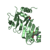
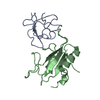
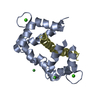
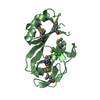
MURAMOYL-PENTAPEPTIDE CARBOXYPEPTIDASE HYDROLYSES INSOLUBLE PEPTIDOGLYCAN CLEAVAGE OF UDP-N- ...MURAMOYL-PENTAPEPTIDE CARBOXYPEPTIDASE HYDROLYSES INSOLUBLE PEPTIDOGLYCAN CLEAVAGE OF UDP-N-ACETYLMURAMOYL-L-ALANYL-D- GAMMA-GLUTAMYL-CARBOXY-L-LYSYL-D-ALANYL-|-D-ALANINE.
Has protein modification
Y
Sequence details
THE FIRST AMINO ACID OF THE NATIVE PROTEIN IS ASP 1 WHICH IS PARTIALLY BLOCKED IN THIS STRUCTURE AS ...THE FIRST AMINO ACID OF THE NATIVE PROTEIN IS ASP 1 WHICH IS PARTIALLY BLOCKED IN THIS STRUCTURE AS A RESULT OF THE CYCLIZATION OF THE FIRST TWO AMINO ACIDS INTO ANHYDROASPARTYLGLYCINE.
-
Experimental details
-
Experiment
Experiment
Method: X-RAY DIFFRACTION
-
Sample preparation
Crystal
Density Matthews: 2.17 Å3/Da / Density % sol: 43.39 %
-
Data collection
Radiation
Scattering type: x-ray
Radiation wavelength
Relative weight: 1
Reflection
Resolution: 1.8→30 Å / Num. obs: 15460 / % possible obs: 100 % / Observed criterion σ(I): 0
-
Processing
Software
Name: PROLSQ / Classification: refinement
Refinement
Resolution: 1.8→6 Å / σ(F): 2 Details: AMINO ACIDS 43 - 109 ARE SIMILAR TO AMINO ACIDS 194 - 261 OF B. SUBTILIS AUTOLYSIN.
Rfactor
Num. reflection
% reflection
Rwork
0.164
-
-
obs
-
12633
84 %
Refinement step
Cycle: LAST / Resolution: 1.8→6 Å
Protein
Nucleic acid
Ligand
Solvent
Total
Num. atoms
1555
0
1
229
1785
Refine LS restraints
Refine-ID
Type
Dev ideal
X-RAY DIFFRACTION
p_bond_d
0.016
X-RAY DIFFRACTION
p_angle_d
0.034
X-RAY DIFFRACTION
p_angle_deg
X-RAY DIFFRACTION
p_planar_d
X-RAY DIFFRACTION
p_hb_or_metal_coord
X-RAY DIFFRACTION
p_mcbond_it
X-RAY DIFFRACTION
p_mcangle_it
X-RAY DIFFRACTION
p_scbond_it
X-RAY DIFFRACTION
p_scangle_it
X-RAY DIFFRACTION
p_plane_restr
X-RAY DIFFRACTION
p_chiral_restr
X-RAY DIFFRACTION
p_singtor_nbd
X-RAY DIFFRACTION
p_multtor_nbd
X-RAY DIFFRACTION
p_xhyhbond_nbd
X-RAY DIFFRACTION
p_xyhbond_nbd
X-RAY DIFFRACTION
p_planar_tor
X-RAY DIFFRACTION
p_staggered_tor
X-RAY DIFFRACTION
p_orthonormal_tor
X-RAY DIFFRACTION
p_transverse_tor
X-RAY DIFFRACTION
p_special_tor
+
About Yorodumi
-
News
-
Feb 9, 2022. New format data for meta-information of EMDB entries
New format data for meta-information of EMDB entries
Version 3 of the EMDB header file is now the official format.
The previous official version 1.9 will be removed from the archive.
In the structure databanks used in Yorodumi, some data are registered as the other names, "COVID-19 virus" and "2019-nCoV". Here are the details of the virus and the list of structure data.
Jan 31, 2019. EMDB accession codes are about to change! (news from PDBe EMDB page)
EMDB accession codes are about to change! (news from PDBe EMDB page)
The allocation of 4 digits for EMDB accession codes will soon come to an end. Whilst these codes will remain in use, new EMDB accession codes will include an additional digit and will expand incrementally as the available range of codes is exhausted. The current 4-digit format prefixed with “EMD-” (i.e. EMD-XXXX) will advance to a 5-digit format (i.e. EMD-XXXXX), and so on. It is currently estimated that the 4-digit codes will be depleted around Spring 2019, at which point the 5-digit format will come into force.
The EM Navigator/Yorodumi systems omit the EMD- prefix.
Related info.:Q: What is EMD? / ID/Accession-code notation in Yorodumi/EM Navigator
Yorodumi is a browser for structure data from EMDB, PDB, SASBDB, etc.
This page is also the successor to EM Navigator detail page, and also detail information page/front-end page for Omokage search.
The word "yorodu" (or yorozu) is an old Japanese word meaning "ten thousand". "mi" (miru) is to see.
Related info.:EMDB / PDB / SASBDB / Comparison of 3 databanks / Yorodumi Search / Aug 31, 2016. New EM Navigator & Yorodumi / Yorodumi Papers / Jmol/JSmol / Function and homology information / Changes in new EM Navigator and Yorodumi
 Movie
Movie Controller


 Open data
 Basic information
 Components
 Keywords
 Function and homology information
 Streptomyces albus (bacteria)
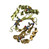
 X-RAY DIFFRACTION / Resolution: 1.8 Å
  Authors
 Citation
 Structure visualization
 Downloads & links
 Other downloads
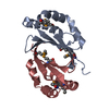


 PDBj
PDBj

 Assembly

 Mass: 65.409 Da / Num. of mol.: 1 / Source method: obtained synthetically / Formula: Zn
 Mass: 18.015 Da / Num. of mol.: 229 / Source method: isolated from a natural source / Formula: H2O
 Sample preparation
 Processing