 Movie
Movie Controller


+ Open data

- Basic information
| Entry | Database: PDB / ID: 1l1f | ||||||
|---|---|---|---|---|---|---|---|
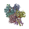
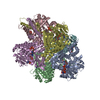
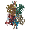
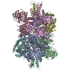
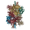
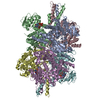
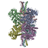
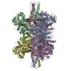
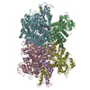
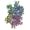
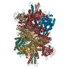
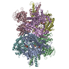
| Title | Structure of human glutamate dehydrogenase-apo form | ||||||
 Components Components | Glutamate Dehydrogenase 1 | ||||||
 Keywords Keywords | OXIDOREDUCTASE / Allostery / negative cooperativity | ||||||
| Function / homology |  Function and homology information Function and homology informationL-glutamate dehydrogenase [NAD(P)+] activity / L-leucine binding / tricarboxylic acid metabolic process / glutamate dehydrogenase [NAD(P)+] / L-glutamate dehydrogenase (NADP+) activity / L-glutamate dehydrogenase (NAD+) activity / Glutamate and glutamine metabolism / L-glutamate catabolic process / : / L-glutamine metabolic process ...L-glutamate dehydrogenase [NAD(P)+] activity / L-leucine binding / tricarboxylic acid metabolic process / glutamate dehydrogenase [NAD(P)+] / L-glutamate dehydrogenase (NADP+) activity / L-glutamate dehydrogenase (NAD+) activity / Glutamate and glutamine metabolism / L-glutamate catabolic process / : / L-glutamine metabolic process / NAD+ binding / substantia nigra development / Mitochondrial protein degradation / Transcriptional activation of mitochondrial biogenesis / ADP binding / positive regulation of insulin secretion / mitochondrial matrix / GTP binding / endoplasmic reticulum / protein homodimerization activity / mitochondrion / ATP binding / cytoplasm Similarity search - Function | ||||||
| Biological species |  Homo sapiens (human) Homo sapiens (human) | ||||||
| Method |  X-RAY DIFFRACTION / MOLECULAR REPLACEMENT / Resolution: 2.7 Å X-RAY DIFFRACTION / MOLECULAR REPLACEMENT / Resolution: 2.7 Å | ||||||
 Authors Authors | Smith, T.J. / Schmidt, T. / Fang, J. / Wu, J. / Siuzdak, G. / Stanley, C.A. | ||||||
 Citation Citation | Journal: J.Mol.Biol. / Year: 2002 Title: The structure of apo human glutamate dehydrogenase details subunit communication and allostery. Authors: Smith, T.J. / Schmidt, T. / Fang, J. / Wu, J. / Siuzdak, G. / Stanley, C.A. | ||||||
| History |
| ||||||
| Remark 400 | COMPOUND NAD binding domain orientation differs among the six chains. |
- Structure visualization
| Structure viewer | Molecule: MolmilJmol/JSmol |
|---|
- Downloads & links
-Download
| PDBx/mmCIF format | 1l1f.cif.gz | 555.3 KB | Display | PDBx/mmCIF format |
|---|---|---|---|---|
| PDB format | pdb1l1f.ent.gz | 465.6 KB | Display | PDB format |
| PDBx/mmJSON format | 1l1f.json.gz | Tree view | PDBx/mmJSON format | |
| Others |  Other downloads Other downloads |
-Validation report
| Arichive directory | https://data.pdbj.org/pub/pdb/validation_reports/l1/1l1fftp://data.pdbj.org/pub/pdb/validation_reports/l1/1l1f | HTTPS FTP |
|---|
-Related structure data
| Related structure data |  1hwx |
|---|---|
| Similar structure data |
-Links
 PDBj
PDBj- Assembly
| Deposited unit | 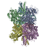
| ||||||||
|---|---|---|---|---|---|---|---|---|---|
| 1 |
| ||||||||
| Unit cell |
|
-Components
| #1: Protein | Mass: 56085.566 Da / Num. of mol.: 6 Source method: isolated from a genetically manipulated source Source: (gene. exp.) Homo sapiens (human) / Production host:  References: UniProt: P00367, glutamate dehydrogenase [NAD(P)+] |
|---|
-Experimental details
-Experiment
| Experiment | Method: X-RAY DIFFRACTION / Number of used crystals: 1 |
|---|
- Sample preparation
| Crystal | Density Matthews: 2.89 Å3/Da / Density % sol: 57.49 % | ||||||||||||||||||||||||||||||||||||||||||||||||||||||||
|---|---|---|---|---|---|---|---|---|---|---|---|---|---|---|---|---|---|---|---|---|---|---|---|---|---|---|---|---|---|---|---|---|---|---|---|---|---|---|---|---|---|---|---|---|---|---|---|---|---|---|---|---|---|---|---|---|---|
| Crystal grow | Temperature: 298 K / Method: vapor diffusion, sitting drop / pH: 7 Details: PEG 8000, NaCl, sodium phosphate, sodium azide, octyl-b-glucopyranoside, methyl pentanediol, pH 7, VAPOR DIFFUSION, SITTING DROP, temperature 298K | ||||||||||||||||||||||||||||||||||||||||||||||||||||||||
| Crystal grow | *PLUS pH: 7 | ||||||||||||||||||||||||||||||||||||||||||||||||||||||||
| Components of the solutions | *PLUS
|
-Data collection
| Diffraction | Mean temperature: 200 K |
|---|---|
| Diffraction source | Source: ROTATING ANODE / Type: RIGAKU RU300 / Wavelength: 1.5418 Å |
| Detector | Type: RIGAKU RAXIS IV / Detector: IMAGE PLATE / Date: Feb 14, 2001 |
| Radiation | Monochromator: graphite / Protocol: SINGLE WAVELENGTH / Monochromatic (M) / Laue (L): M / Scattering type: x-ray |
| Radiation wavelength | Wavelength: 1.5418 Å / Relative weight: 1 |
| Reflection | Resolution: 2.7→30 Å / Num. all: 94574 / Num. obs: 91356 / % possible obs: 90.5 % / Observed criterion σ(F): 1 / Observed criterion σ(I): 1 / Redundancy: 2.1 % / Rsym value: 0.047 |
| Reflection | *PLUS Lowest resolution: 30 Å / Num. obs: 94574 / Rmerge(I) obs: 0.047 |
| Reflection shell | *PLUS Highest resolution: 2.7 Å / Lowest resolution: 2.82 Å / % possible obs: 54.8 % / Num. unique obs: 7168 / Rmerge(I) obs: 0.216 / Mean I/σ(I) obs: 2.2 |
- Processing
| Software |
| ||||||||||||||||||||
|---|---|---|---|---|---|---|---|---|---|---|---|---|---|---|---|---|---|---|---|---|---|
| Refinement | Method to determine structure: MOLECULAR REPLACEMENT / Resolution: 2.7→8 Å / σ(F): 2 / Stereochemistry target values: Engh & Huber
| ||||||||||||||||||||
| Refinement step | Cycle: LAST / Resolution: 2.7→8 Å
| ||||||||||||||||||||
| Software | *PLUS Name: 0 / Classification: refinement | ||||||||||||||||||||
| Refinement | *PLUS Highest resolution: 2.7 Å / Lowest resolution: 8 Å / σ(F): 2 / Num. reflection Rfree: 30 / % reflection Rfree: 5 % / Rfactor obs: 0.262 | ||||||||||||||||||||
| Solvent computation | *PLUS | ||||||||||||||||||||
| Displacement parameters | *PLUS |
