 Movie
Movie Controller

[English] 日本語
 Yorodumi
- PDB-1kzo: PROTEIN FARNESYLTRANSFERASE COMPLEXED WITH FARNESYLATED K-RAS4B P... -
+ Open data

- Basic information
| Entry | Database: PDB / ID: 1kzo | ||||||
|---|---|---|---|---|---|---|---|
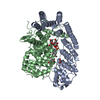
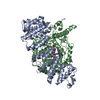
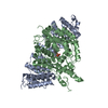
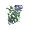
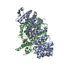
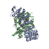
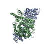
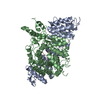
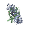
| Title | PROTEIN FARNESYLTRANSFERASE COMPLEXED WITH FARNESYLATED K-RAS4B PEPTIDE PRODUCT AND FARNESYL DIPHOSPHATE SUBSTRATE BOUND SIMULTANEOUSLY | ||||||
 Components Components |
| ||||||
 Keywords Keywords | TRANSFERASE/TRANSFERASE SUBSTRATE / FTASE / PFT / PFTASE / FT / FPT / FARNESYLTRANSFERASE / FARNESYL TRANSFERASE / FARNESYL PROTEIN TRANSFERASE / CAAX / RAS / CANCER / PRODUCT / SUBSTRATE / TRANSFERASE-TRANSFERASE SUBSTRATE COMPLEX | ||||||
| Function / homology |  Function and homology information Function and homology informationApoptotic cleavage of cellular proteins / Inactivation, recovery and regulation of the phototransduction cascade / RAS processing / protein geranylgeranyltransferase activity / peptide pheromone maturation / protein geranylgeranyltransferase type I / CAAX-protein geranylgeranyltransferase activity / CAAX-protein geranylgeranyltransferase complex / protein farnesylation / protein farnesyltransferase ...Apoptotic cleavage of cellular proteins / Inactivation, recovery and regulation of the phototransduction cascade / RAS processing / protein geranylgeranyltransferase activity / peptide pheromone maturation / protein geranylgeranyltransferase type I / CAAX-protein geranylgeranyltransferase activity / CAAX-protein geranylgeranyltransferase complex / protein farnesylation / protein farnesyltransferase / protein farnesyltransferase activity / protein farnesyltransferase complex / Rab geranylgeranyltransferase activity / protein geranylgeranylation / regulation of fibroblast proliferation / nuclear envelope organization / geranylgeranyl diphosphate synthase activity / acetyltransferase activator activity / positive regulation of skeletal muscle acetylcholine-gated channel clustering / positive regulation of Ras protein signal transduction / microtubule associated complex / response to mineralocorticoid / enzyme-linked receptor protein signaling pathway / GMP binding / forebrain astrocyte development / LRR domain binding / regulation of synaptic transmission, GABAergic / negative regulation of epithelial cell differentiation / response to isolation stress / regulation of microtubule-based movement / response to gravity / epithelial tube branching involved in lung morphogenesis / type I pneumocyte differentiation / Rac protein signal transduction / myoblast proliferation / Signaling by RAS GAP mutants / Signaling by RAS GTPase mutants / Activation of RAS in B cells / RAS signaling downstream of NF1 loss-of-function variants / RUNX3 regulates p14-ARF / skeletal muscle cell differentiation / positive regulation of glial cell proliferation / SOS-mediated signalling / Activated NTRK3 signals through RAS / Activated NTRK2 signals through RAS / cardiac muscle cell proliferation / SHC1 events in ERBB4 signaling / Signalling to RAS / alpha-tubulin binding / SHC-related events triggered by IGF1R / Activated NTRK2 signals through FRS2 and FRS3 / Estrogen-stimulated signaling through PRKCZ / positive regulation of Rac protein signal transduction / SHC-mediated cascade:FGFR3 / glial cell proliferation / MET activates RAS signaling / SHC-mediated cascade:FGFR2 / SHC-mediated cascade:FGFR4 / Signaling by PDGFRA transmembrane, juxtamembrane and kinase domain mutants / Signaling by PDGFRA extracellular domain mutants / PTK6 Regulates RHO GTPases, RAS GTPase and MAP kinases / Erythropoietin activates RAS / SHC-mediated cascade:FGFR1 / Signaling by FGFR4 in disease / Signaling by CSF3 (G-CSF) / FRS-mediated FGFR3 signaling / Signaling by FLT3 ITD and TKD mutants / FRS-mediated FGFR2 signaling / FRS-mediated FGFR4 signaling / p38MAPK events / striated muscle cell differentiation / FRS-mediated FGFR1 signaling / Signaling by FGFR3 in disease / Tie2 Signaling / protein-membrane adaptor activity / Signaling by FGFR2 in disease / positive regulation of cell cycle / Signaling by FLT3 fusion proteins / GRB2 events in EGFR signaling / SHC1 events in EGFR signaling / FLT3 Signaling / Signaling by FGFR1 in disease / EGFR Transactivation by Gastrin / NCAM signaling for neurite out-growth / CD209 (DC-SIGN) signaling / Downstream signal transduction / GRB2 events in ERBB2 signaling / homeostasis of number of cells within a tissue / Insulin receptor signalling cascade / SHC1 events in ERBB2 signaling / Constitutive Signaling by Overexpressed ERBB2 / Signaling by phosphorylated juxtamembrane, extracellular and kinase domain KIT mutants / Ras activation upon Ca2+ influx through NMDA receptor / response to glucocorticoid / VEGFR2 mediated cell proliferation / small monomeric GTPase / lipid metabolic process / FCERI mediated MAPK activation / wound healing / liver development Similarity search - Function | ||||||
| Biological species |  | ||||||
| Method |  X-RAY DIFFRACTION / MOLECULAR REPLACEMENT / Resolution: 2.2 Å X-RAY DIFFRACTION / MOLECULAR REPLACEMENT / Resolution: 2.2 Å | ||||||
 Authors Authors | Long, S.B. / Casey, P.J. / Beese, L.S. | ||||||
 Citation Citation | Journal: Nature / Year: 2002 Title: The Reaction Path of Protein Farnesyltransferase at Atomic Resolution Authors: Long, S.B. / Casey, P.J. / Beese, L.S. #1: Journal: Structure / Year: 2000Title: The Basis for K-Ras4B Binding Specificity to Protein Farnesyltransferase Revealed by 2A Resolution Ternary Complex Structures Authors: Long, S.B. / Casey, P.J. / Beese, L.S. #2: Journal: Science / Year: 1997Title: Crystal Structure of Protein Farnesyltransferase at 2.25A Resolution Authors: Park, H.W. / Boduluri, S.R. / Moomaw, J.F. / Casey, P.J. / Beese, L.S. #3: Journal: Biochemistry / Year: 1998Title: Co-Crystal Structure of Mammalian Protein Farnesyltransferase with a Farnesyl Diphosphate Substrate Authors: Long, S.B. / Casey, P.J. / Beese, L.S. | ||||||
| History |
|
- Structure visualization
| Structure viewer | Molecule: MolmilJmol/JSmol |
|---|
- Downloads & links
-Download
| PDBx/mmCIF format | 1kzo.cif.gz | 174.7 KB | Display | PDBx/mmCIF format |
|---|---|---|---|---|
| PDB format | pdb1kzo.ent.gz | 134.3 KB | Display | PDB format |
| PDBx/mmJSON format | 1kzo.json.gz | Tree view | PDBx/mmJSON format | |
| Others |  Other downloads Other downloads |
-Validation report
| Arichive directory | https://data.pdbj.org/pub/pdb/validation_reports/kz/1kzoftp://data.pdbj.org/pub/pdb/validation_reports/kz/1kzo | HTTPS FTP |
|---|
-Related structure data
| Related structure data |  1kzpC  1d8dS C: citing same article ( S: Starting model for refinement |
|---|---|
| Similar structure data |


-Links
 PDBj
PDBj


- Assembly
| Deposited unit | 
| ||||||||
|---|---|---|---|---|---|---|---|---|---|
| 1 |
| ||||||||
| Unit cell |
| ||||||||
| Details | The biological unit of protein farnesyltransferase (FTase) is a heterodimer of alpha and beta subunits. There is one FTase heterodimer per asymmetric unit. |
-Components
-Protein Farnesyltransferase ... , 2 types, 2 molecules AB
| #1: Protein | Mass: 44098.145 Da / Num. of mol.: 1 Source method: isolated from a genetically manipulated source Source: (gene. exp.)   Spodoptera frugiperda (fall armyworm) / References: UniProt: Q04631, squalene synthase Spodoptera frugiperda (fall armyworm) / References: UniProt: Q04631, squalene synthase |
|---|---|
| #2: Protein | Mass: 48722.281 Da / Num. of mol.: 1 Source method: isolated from a genetically manipulated source Source: (gene. exp.) Spodoptera frugiperda (fall armyworm) / References: UniProt: Q02293, squalene synthase |
-Protein/peptide , 1 types, 1 molecules C
| #3: Protein/peptide | Mass: 1298.723 Da / Num. of mol.: 1 / Source method: obtained synthetically Details: THE PEPTIDE WAS CHEMICALLY SYNTHESIZED. THE SEQUENCE OF THE PEPTIDE IS NATURALLY FOUND IN HOMO SAPIENS. References: UniProt: P01118, UniProt: P01116*PLUS |
|---|
-Non-polymers , 5 types, 432 molecules 


| #4: Chemical | ChemComp-ZN /  Mass: 65.409 Da / Num. of mol.: 1 / Source method: obtained synthetically / Formula: Zn Mass: 65.409 Da / Num. of mol.: 1 / Source method: obtained synthetically / Formula: Zn |
|---|---|
| #5: Chemical | ChemComp-FPP /  Mass: 382.326 Da / Num. of mol.: 1 / Source method: obtained synthetically / Formula: C15H28O7P2 Mass: 382.326 Da / Num. of mol.: 1 / Source method: obtained synthetically / Formula: C15H28O7P2 |
| #6: Chemical | ChemComp-ACY /  Mass: 60.052 Da / Num. of mol.: 1 / Source method: obtained synthetically / Formula: C2H4O2 Mass: 60.052 Da / Num. of mol.: 1 / Source method: obtained synthetically / Formula: C2H4O2 |
| #7: Chemical | ChemComp-FAR /  Mass: 206.367 Da / Num. of mol.: 1 / Source method: obtained synthetically / Formula: C15H26 Mass: 206.367 Da / Num. of mol.: 1 / Source method: obtained synthetically / Formula: C15H26 |
| #8: Water | ChemComp-HOH / Mass: 18.015 Da / Num. of mol.: 428 / Source method: isolated from a natural source / Formula: H2O |
-Details
| Has protein modification | Y |
|---|
-Experimental details
-Experiment
| Experiment | Method: X-RAY DIFFRACTION / Number of used crystals: 1 |
|---|
- Sample preparation
| Crystal | Density Matthews: 3.12 Å3/Da / Density % sol: 60.57 % | ||||||||||||||||||||
|---|---|---|---|---|---|---|---|---|---|---|---|---|---|---|---|---|---|---|---|---|---|
| Crystal grow | Temperature: 290 K / Method: vapor diffusion, hanging drop / pH: 5.7 Details: PEG 8000, Ammonium Acetate, Magnesium Chloride, DTT, Tris-HCl, pH 5.70, VAPOR DIFFUSION, HANGING DROP, temperature 290K | ||||||||||||||||||||
| Crystal grow | *PLUS Temperature: 17 ℃ / pH: 5.7 / Details: Long, S.B., (2000) Structure, 8, 209. | ||||||||||||||||||||
| Components of the solutions | *PLUS
|
-Data collection
| Diffraction | Mean temperature: 100 K |
|---|---|
| Diffraction source | Source: ROTATING ANODE / Type: RIGAKU RUH3R / Wavelength: 1.5418 / Wavelength: 1.5418 Å |
| Detector | Type: RIGAKU RAXIS IV / Detector: IMAGE PLATE / Date: Mar 7, 2000 / Details: Mirrors |
| Radiation | Monochromator: Ni Filter / Protocol: SINGLE WAVELENGTH / Monochromatic (M) / Laue (L): M / Scattering type: x-ray |
| Radiation wavelength | Wavelength: 1.5418 Å / Relative weight: 1 |
| Reflection | Resolution: 2.2→50 Å / Num. obs: 56209 / % possible obs: 96 % / Observed criterion σ(I): -3 / Redundancy: 4.51 % / Biso Wilson estimate: 32.8 Å2 / Rsym value: 0.089 / Net I/σ(I): 16.1 |
| Reflection shell | Resolution: 2.2→2.23 Å / Redundancy: 3.2 % / Mean I/σ(I) obs: 1.8 / Num. unique all: 1705 / Rsym value: 0.63 / % possible all: 87.9 |
| Reflection | *PLUS Highest resolution: 2.2 Å / Lowest resolution: 50 Å / Num. measured all: 253335 / Rmerge(I) obs: 0.089 |
| Reflection shell | *PLUS Highest resolution: 2.2 Å / Lowest resolution: 2.3 Å / Rmerge(I) obs: 0.56 |
- Processing
| Software |
| ||||||||||||||||||||||||||||||||||||
|---|---|---|---|---|---|---|---|---|---|---|---|---|---|---|---|---|---|---|---|---|---|---|---|---|---|---|---|---|---|---|---|---|---|---|---|---|---|
| Refinement | Method to determine structure: MOLECULAR REPLACEMENT Starting model: PDB entry 1D8D Resolution: 2.2→50 Å / Rfactor Rfree error: 0.004 / Data cutoff high absF: 10000000 / Data cutoff low absF: 0 / Isotropic thermal model: RESTRAINED / Cross valid method: THROUGHOUT / σ(F): 2 / Stereochemistry target values: Engh & Huber
| ||||||||||||||||||||||||||||||||||||
| Displacement parameters | Biso mean: 34.9 Å2
| ||||||||||||||||||||||||||||||||||||
| Refine analyze |
| ||||||||||||||||||||||||||||||||||||
| Refinement step | Cycle: LAST / Resolution: 2.2→50 Å
| ||||||||||||||||||||||||||||||||||||
| Refine LS restraints |
| ||||||||||||||||||||||||||||||||||||
| LS refinement shell | Resolution: 2.2→2.3 Å / Rfactor Rfree error: 0.019 / Total num. of bins used: 8
| ||||||||||||||||||||||||||||||||||||
| Xplor file |
| ||||||||||||||||||||||||||||||||||||
| Refinement | *PLUS Highest resolution: 2.2 Å / Lowest resolution: 50 Å / % reflection Rfree: 5 % / Rfactor all: 0.162 / Rfactor obs: 0.159 / Rfactor Rfree: 0.204 / Rfactor Rwork: 0.16 | ||||||||||||||||||||||||||||||||||||
| Solvent computation | *PLUS | ||||||||||||||||||||||||||||||||||||
| Displacement parameters | *PLUS | ||||||||||||||||||||||||||||||||||||
| Refine LS restraints | *PLUS
| ||||||||||||||||||||||||||||||||||||
| LS refinement shell | *PLUS Highest resolution: 2.2 Å / Lowest resolution: 2.3 Å / Rfactor Rfree: 0.279 / Rfactor Rwork: 0.255 |
