 Movie
Movie Controller

[English] 日本語
 Yorodumi
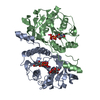
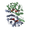
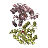
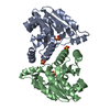
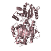
- PDB-1kws: CRYSTAL STRUCTURE OF BETA1,3-GLUCURONYLTRANSFERASE I IN COMPLEX W... -
+ Open data

- Basic information
| Entry | Database: PDB / ID: 1kws | ||||||
|---|---|---|---|---|---|---|---|
| Title | CRYSTAL STRUCTURE OF BETA1,3-GLUCURONYLTRANSFERASE I IN COMPLEX WITH THE ACTIVE UDP-GLCUA DONOR | ||||||
 Components Components | BETA-1,3-GLUCURONYLTRANSFERASE 3 | ||||||
 Keywords Keywords | TRANSFERASE / DXD / NTP binding domain | ||||||
| Function / homology |  Function and homology information Function and homology informationdermatan sulfate proteoglycan biosynthetic process / galactosylgalactosylxylosylprotein 3-beta-glucuronosyltransferase / galactosylgalactosylxylosylprotein 3-beta-glucuronosyltransferase activity / glucuronosyltransferase activity / Defective B3GAT3 causes JDSSDHD / Glycosaminoglycan-protein linkage region biosynthesis / glycosaminoglycan biosynthetic process / chondroitin sulfate proteoglycan biosynthetic process / heparan sulfate proteoglycan biosynthetic process / positive regulation of catalytic activity ...dermatan sulfate proteoglycan biosynthetic process / galactosylgalactosylxylosylprotein 3-beta-glucuronosyltransferase / galactosylgalactosylxylosylprotein 3-beta-glucuronosyltransferase activity / glucuronosyltransferase activity / Defective B3GAT3 causes JDSSDHD / Glycosaminoglycan-protein linkage region biosynthesis / glycosaminoglycan biosynthetic process / chondroitin sulfate proteoglycan biosynthetic process / heparan sulfate proteoglycan biosynthetic process / positive regulation of catalytic activity / positive regulation of intracellular protein transport / cis-Golgi network / protein phosphatase activator activity / carbohydrate metabolic process / Golgi membrane / Golgi apparatus / extracellular exosome / membrane / metal ion binding Similarity search - Function | ||||||
| Biological species |  Homo sapiens (human) Homo sapiens (human) | ||||||
| Method |  X-RAY DIFFRACTION / FOURIER SYNTHESIS / Resolution: 2.1 Å X-RAY DIFFRACTION / FOURIER SYNTHESIS / Resolution: 2.1 Å | ||||||
 Authors Authors | Pedersen, L.C. / Darden, T.A. / Negishi, M. | ||||||
 Citation Citation | Journal: J.Biol.Chem. / Year: 2002 Title: Crystal structure of beta 1,3-glucuronyltransferase I in complex with active donor substrate UDP-GlcUA. Authors: Pedersen, L.C. / Darden, T.A. / Negishi, M. | ||||||
| History |
|
- Structure visualization
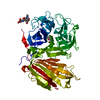
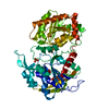
| Structure viewer | Molecule: MolmilJmol/JSmol |
|---|
- Downloads & links
-Download
| PDBx/mmCIF format | 1kws.cif.gz | 119.8 KB | Display | PDBx/mmCIF format |
|---|---|---|---|---|
| PDB format | pdb1kws.ent.gz | 90.2 KB | Display | PDB format |
| PDBx/mmJSON format | 1kws.json.gz | Tree view | PDBx/mmJSON format | |
| Others |  Other downloads Other downloads |
-Validation report
| Arichive directory | https://data.pdbj.org/pub/pdb/validation_reports/kw/1kwsftp://data.pdbj.org/pub/pdb/validation_reports/kw/1kws | HTTPS FTP |
|---|
-Related structure data
| Related structure data |  1fggS S: Starting model for refinement |
|---|---|
| Similar structure data |


-Links
 PDBj
PDBj

- Assembly
| Deposited unit | 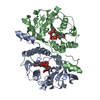
| ||||||||
|---|---|---|---|---|---|---|---|---|---|
| 1 |
| ||||||||
| Unit cell |
|
-Components
| #1: Protein | Mass: 28992.064 Da / Num. of mol.: 2 / Fragment: Residue 76-335 Source method: isolated from a genetically manipulated source Source: (gene. exp.) Homo sapiens (human) / Plasmid: PET28A / Species (production host): Escherichia coli / Production host:  #2: Chemical |   Mass: 54.938 Da / Num. of mol.: 2 / Source method: obtained synthetically / Formula: Mn Mass: 54.938 Da / Num. of mol.: 2 / Source method: obtained synthetically / Formula: Mn#3: Chemical | 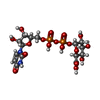  Mass: 580.285 Da / Num. of mol.: 2 / Source method: obtained synthetically / Formula: C15H22N2O18P2 Mass: 580.285 Da / Num. of mol.: 2 / Source method: obtained synthetically / Formula: C15H22N2O18P2#4: Water | ChemComp-HOH / |  Mass: 18.015 Da / Num. of mol.: 359 / Source method: isolated from a natural source / Formula: H2O Mass: 18.015 Da / Num. of mol.: 359 / Source method: isolated from a natural source / Formula: H2O |
|---|
-Experimental details
-Experiment
| Experiment | Method: X-RAY DIFFRACTION / Number of used crystals: 1 |
|---|
- Sample preparation
| Crystal | Density Matthews: 2.43 Å3/Da / Density % sol: 49.38 % | ||||||||||||||||||||||||||||||||||||||||||||||||||||||||
|---|---|---|---|---|---|---|---|---|---|---|---|---|---|---|---|---|---|---|---|---|---|---|---|---|---|---|---|---|---|---|---|---|---|---|---|---|---|---|---|---|---|---|---|---|---|---|---|---|---|---|---|---|---|---|---|---|---|
| Crystal grow | Temperature: 296 K / Method: vapor diffusion, hanging drop / pH: 6 Details: MES, MME-PEG2K, pH 6.0, VAPOR DIFFUSION, HANGING DROP, temperature 296K | ||||||||||||||||||||||||||||||||||||||||||||||||||||||||
| Crystal grow | *PLUS Temperature: 23 ℃ / pH: 7.5 | ||||||||||||||||||||||||||||||||||||||||||||||||||||||||
| Components of the solutions | *PLUS
|
-Data collection
| Diffraction | Mean temperature: 103 K |
|---|---|
| Diffraction source | Source: ROTATING ANODE / Type: RIGAKU RU300 / Wavelength: 1.5418 Å |
| Detector | Type: RIGAKU RAXIS IV / Detector: IMAGE PLATE / Date: Aug 9, 2000 / Details: Yale Mirrors |
| Radiation | Monochromator: Yale Mirrors / Protocol: SINGLE WAVELENGTH / Monochromatic (M) / Laue (L): M / Scattering type: x-ray |
| Radiation wavelength | Wavelength: 1.5418 Å / Relative weight: 1 |
| Reflection | Resolution: 2.1→25 Å / Num. all: 32779 / Num. obs: 32764 / % possible obs: 99.5 % / Observed criterion σ(I): -3 / Redundancy: 3.4 % / Biso Wilson estimate: 7.9 Å2 / Rmerge(I) obs: 0.082 / Net I/σ(I): 12.8 |
| Reflection shell | Resolution: 2.1→2.18 Å / Redundancy: 2.67 % / Rmerge(I) obs: 0.309 / Mean I/σ(I) obs: 3 / Num. unique all: 3134 / % possible all: 96 |
| Reflection | *PLUS Num. obs: 32779 / Num. measured all: 112504 / Rmerge(I) obs: 0.082 |
| Reflection shell | *PLUS % possible obs: 96 % / Rmerge(I) obs: 0.309 |
- Processing
| Software |
| ||||||||||||||||||||||||||||||||||||
|---|---|---|---|---|---|---|---|---|---|---|---|---|---|---|---|---|---|---|---|---|---|---|---|---|---|---|---|---|---|---|---|---|---|---|---|---|---|
| Refinement | Method to determine structure: FOURIER SYNTHESIS Starting model: PDB ENTRY 1FGG Resolution: 2.1→24.68 Å / Rfactor Rfree error: 0.006 / Data cutoff high absF: 139032.36 / Data cutoff low absF: 0 / Isotropic thermal model: RESTRAINED / Cross valid method: THROUGHOUT / σ(F): 0 / Stereochemistry target values: Engh & Huber
| ||||||||||||||||||||||||||||||||||||
| Solvent computation | Solvent model: FLAT MODEL / Bsol: 41.4552 Å2 / ksol: 0.351798 e/Å3 | ||||||||||||||||||||||||||||||||||||
| Displacement parameters | Biso mean: 22.2 Å2
| ||||||||||||||||||||||||||||||||||||
| Refine analyze |
| ||||||||||||||||||||||||||||||||||||
| Refinement step | Cycle: LAST / Resolution: 2.1→24.68 Å
| ||||||||||||||||||||||||||||||||||||
| Refine LS restraints |
| ||||||||||||||||||||||||||||||||||||
| LS refinement shell | Resolution: 2.1→2.23 Å / Rfactor Rfree error: 0.016 / Total num. of bins used: 6
| ||||||||||||||||||||||||||||||||||||
| Xplor file |
| ||||||||||||||||||||||||||||||||||||
| Refinement | *PLUS Highest resolution: 2.1 Å / Lowest resolution: 50 Å / % reflection Rfree: 5 % / Rfactor all: 0.188 / Rfactor obs: 0.187 / Rfactor Rfree: 0.223 / Rfactor Rwork: 0.187 | ||||||||||||||||||||||||||||||||||||
| Solvent computation | *PLUS | ||||||||||||||||||||||||||||||||||||
| Displacement parameters | *PLUS | ||||||||||||||||||||||||||||||||||||
| Refine LS restraints | *PLUS
| ||||||||||||||||||||||||||||||||||||
| LS refinement shell | *PLUS Rfactor Rfree: 0.262 / Rfactor Rwork: 0.22 / Rfactor obs: 0.22 |
