 Movie
Movie Controller

[English] 日本語
 Yorodumi
- PDB-1kuq: CRYSTAL STRUCTURE OF T3C MUTANT S15 RIBOSOMAL PROTEIN IN COMPLEX ... -
+ Open data

- Basic information
| Entry | Database: PDB / ID: 1kuq | ||||||
|---|---|---|---|---|---|---|---|
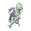
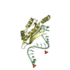
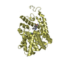
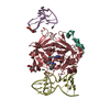
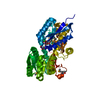
| Title | CRYSTAL STRUCTURE OF T3C MUTANT S15 RIBOSOMAL PROTEIN IN COMPLEX WITH 16S RRNA | ||||||
 Components Components |
| ||||||
 Keywords Keywords | RIBOSOME / rRNA-protein complex | ||||||
| Function / homology |  Function and homology information Function and homology informationcytosolic small ribosomal subunit / rRNA binding / structural constituent of ribosome / translation Similarity search - Function | ||||||
| Biological species |   Thermus thermophilus (bacteria) Thermus thermophilus (bacteria) | ||||||
| Method |  X-RAY DIFFRACTION / SYNCHROTRON / MOLECULAR REPLACEMENT / Resolution: 2.84 Å X-RAY DIFFRACTION / SYNCHROTRON / MOLECULAR REPLACEMENT / Resolution: 2.84 Å | ||||||
 Authors Authors | Nikulin, A.D. / Tishchenko, S. / Revtovich, S. / Ehresmann, B. / Ehresmann, C. / Dumas, P. / Garber, M. / Nikonov, S. / Nevskaya, N. | ||||||
 Citation Citation | Journal: Biochemistry Mosc. / Year: 2004 Title: Role of N-terminal helix in interaction of ribosomal protein S15 with 16S rRNA. Authors: Revtovich, S.V. / Nikulin, A.D. / Nikonov, S.V. #1: Journal: Nat.Struct.Biol. / Year: 2000Title: Crystal structure of the S15-rRNA complex Authors: Nikulin, A. / Serganov, A. / Ennifar, E. / Tishchenko, S. / Nevskaya, N. / Shepard, W. / Portier, C. / Garber, M. / Ehresmann, B. / Ehresmann, C. / Nikonov, S. / Dumas, P. | ||||||
| History |
|
- Structure visualization
| Structure viewer | Molecule: MolmilJmol/JSmol |
|---|
- Downloads & links
-Download
| PDBx/mmCIF format | 1kuq.cif.gz | 65.1 KB | Display | PDBx/mmCIF format |
|---|---|---|---|---|
| PDB format | pdb1kuq.ent.gz | 45.3 KB | Display | PDB format |
| PDBx/mmJSON format | 1kuq.json.gz | Tree view | PDBx/mmJSON format | |
| Others |  Other downloads Other downloads |
-Validation report
| Arichive directory | https://data.pdbj.org/pub/pdb/validation_reports/ku/1kuqftp://data.pdbj.org/pub/pdb/validation_reports/ku/1kuq | HTTPS FTP |
|---|
-Related structure data
| Related structure data |  1dk1S S: Starting model for refinement |
|---|---|
| Similar structure data |


-Links
 PDBj
PDBj


- Assembly
| Deposited unit | 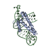
| ||||||||
|---|---|---|---|---|---|---|---|---|---|
| 1 |
| ||||||||
| Unit cell |
|
-Components
| #1: RNA chain | Mass: 18464.963 Da / Num. of mol.: 1 Source method: isolated from a genetically manipulated source Source: (gene. exp.) Thermus thermophilus (bacteria) / Production host: |
|---|---|
| #2: Protein | Mass: 10366.200 Da / Num. of mol.: 1 / Mutation: T3C Source method: isolated from a genetically manipulated source Source: (gene. exp.) Thermus thermophilus (bacteria) / Production host: |
| #3: Chemical | ChemComp-SO4 /   Mass: 96.063 Da / Num. of mol.: 1 / Source method: obtained synthetically / Formula: SO4 Mass: 96.063 Da / Num. of mol.: 1 / Source method: obtained synthetically / Formula: SO4 |
| #4: Water | ChemComp-HOH /  Mass: 18.015 Da / Num. of mol.: 6 / Source method: isolated from a natural source / Formula: H2O Mass: 18.015 Da / Num. of mol.: 6 / Source method: isolated from a natural source / Formula: H2O |
-Experimental details
-Experiment
| Experiment | Method: X-RAY DIFFRACTION / Number of used crystals: 1 |
|---|
- Sample preparation
| Crystal | Density Matthews: 2.67 Å3/Da / Density % sol: 53.98 % | ||||||||||||||||||||
|---|---|---|---|---|---|---|---|---|---|---|---|---|---|---|---|---|---|---|---|---|---|
| Crystal grow | Temperature: 295 K / Method: vapor diffusion, hanging drop / pH: 6.2 Details: ammonium sulphate, KCl, MgCl2, sodium cacodylate, pH 6.2, VAPOR DIFFUSION, HANGING DROP, temperature 295K | ||||||||||||||||||||
| Components of the solutions |
|
-Data collection
| Diffraction | Mean temperature: 100 K |
|---|---|
| Diffraction source | Source: SYNCHROTRON / Site: ESRF  / Beamline: ID14-2 / Wavelength: 0.933 Å / Beamline: ID14-2 / Wavelength: 0.933 Å |
| Detector | Type: MARRESEARCH / Detector: CCD / Date: Sep 21, 1999 |
| Radiation | Protocol: SINGLE WAVELENGTH / Monochromatic (M) / Laue (L): M / Scattering type: x-ray |
| Radiation wavelength | Wavelength: 0.933 Å / Relative weight: 1 |
| Reflection | Resolution: 2.84→25 Å / Num. all: 7828 / Num. obs: 7828 / % possible obs: 99.8 % / Observed criterion σ(F): 0 / Observed criterion σ(I): 0 / Redundancy: 5.6 % / Biso Wilson estimate: 117.9 Å2 / Rmerge(I) obs: 0.049 |
| Reflection shell | Resolution: 2.84→2.92 Å / Rmerge(I) obs: 0.12 / % possible all: 99.9 |
- Processing
| Software |
| ||||||||||||||||||||||||||||||||||||||||||||||||||||||||||||||||||||||||||||||||
|---|---|---|---|---|---|---|---|---|---|---|---|---|---|---|---|---|---|---|---|---|---|---|---|---|---|---|---|---|---|---|---|---|---|---|---|---|---|---|---|---|---|---|---|---|---|---|---|---|---|---|---|---|---|---|---|---|---|---|---|---|---|---|---|---|---|---|---|---|---|---|---|---|---|---|---|---|---|---|---|---|---|
| Refinement | Method to determine structure: MOLECULAR REPLACEMENT Starting model: PDB entry 1DK1 Resolution: 2.84→24.41 Å / Rfactor Rfree error: 0.016 / Data cutoff high absF: 1157536.51 / Data cutoff low absF: 0 / Isotropic thermal model: RESTRAINED / Cross valid method: THROUGHOUT / σ(F): 2 / Stereochemistry target values: Engh & Huber
| ||||||||||||||||||||||||||||||||||||||||||||||||||||||||||||||||||||||||||||||||
| Solvent computation | Solvent model: FLAT MODEL / Bsol: 69.8947 Å2 / ksol: 0.362129 e/Å3 | ||||||||||||||||||||||||||||||||||||||||||||||||||||||||||||||||||||||||||||||||
| Displacement parameters | Biso mean: 56.9 Å2
| ||||||||||||||||||||||||||||||||||||||||||||||||||||||||||||||||||||||||||||||||
| Refine analyze |
| ||||||||||||||||||||||||||||||||||||||||||||||||||||||||||||||||||||||||||||||||
| Refinement step | Cycle: LAST / Resolution: 2.84→24.41 Å
| ||||||||||||||||||||||||||||||||||||||||||||||||||||||||||||||||||||||||||||||||
| Refine LS restraints |
| ||||||||||||||||||||||||||||||||||||||||||||||||||||||||||||||||||||||||||||||||
| LS refinement shell | Resolution: 2.84→3.02 Å / Rfactor Rfree error: 0.06 / Total num. of bins used: 6
| ||||||||||||||||||||||||||||||||||||||||||||||||||||||||||||||||||||||||||||||||
| Xplor file |
|
