 Movie
Movie Controller


+ Open data

- Basic information
| Entry | Database: PDB / ID: 1ktn | ||||||
|---|---|---|---|---|---|---|---|
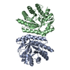
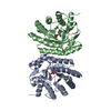
| Title | Structural Genomics, Protein EC1535 | ||||||
 Components Components | 2-deoxyribose-5-phosphate aldolase | ||||||
 Keywords Keywords | STRUCTURAL GENOMICS / LYASE / BETA BARREL / PSI / Protein Structure Initiative / Midwest Center for Structural Genomics / MCSG | ||||||
| Function / homology |  Function and homology information Function and homology informationdeoxyribose-phosphate aldolase / deoxyribose-phosphate aldolase activity / 2-deoxyribose 1-phosphate catabolic process / deoxyribonucleotide catabolic process / nucleobase-containing small molecule interconversion / carbohydrate catabolic process / lyase activity / DNA damage response / membrane / cytosol Similarity search - Function | ||||||
| Biological species |  | ||||||
| Method |  X-RAY DIFFRACTION / SYNCHROTRON / MAD / Resolution: 1.4 Å X-RAY DIFFRACTION / SYNCHROTRON / MAD / Resolution: 1.4 Å | ||||||
 Authors Authors | Zhang, R. / Joachimiak, A. / Edwards, A. / Skarina, T. / Evdokimova, E. / Savchenko, A. / Midwest Center for Structural Genomics (MCSG) | ||||||
 Citation Citation | Journal: To be Published Title: The 1.5A crystal structure of 2-deoxyribose-5-phosphate aldlase Authors: Zhang, R. / Joachimiak, A. / Edwards, A. / Skarina, T. / Evdokimova, E. / Savchenko, A. | ||||||
| History |
|
- Structure visualization
| Structure viewer | Molecule: MolmilJmol/JSmol |
|---|
- Downloads & links
-Download
| PDBx/mmCIF format | 1ktn.cif.gz | 115.2 KB | Display | PDBx/mmCIF format |
|---|---|---|---|---|
| PDB format | pdb1ktn.ent.gz | 89.1 KB | Display | PDB format |
| PDBx/mmJSON format | 1ktn.json.gz | Tree view | PDBx/mmJSON format | |
| Others |  Other downloads Other downloads |
-Validation report
| Arichive directory | https://data.pdbj.org/pub/pdb/validation_reports/kt/1ktnftp://data.pdbj.org/pub/pdb/validation_reports/kt/1ktn | HTTPS FTP |
|---|
-Related structure data
| Similar structure data | |
|---|---|
| Other databases |


-Links
 PDBj
PDBj
- Assembly
| Deposited unit | 
| ||||||||
|---|---|---|---|---|---|---|---|---|---|
| 1 |
| ||||||||
| Unit cell |
| ||||||||
| Details | EC1535 existed as monomer. Chain A and Chain B represent two molecules in asymmetric unit. |
-Components
| #1: Protein | Mass: 26914.955 Da / Num. of mol.: 2 Source method: isolated from a genetically manipulated source Source: (gene. exp.) Plasmid details: T7 promoter system in BL21(DE3) E. coli strain Species (production host): Escherichia coli / Production host: #2: Water | ChemComp-HOH / |  Mass: 18.015 Da / Num. of mol.: 631 / Source method: isolated from a natural source / Formula: H2O Mass: 18.015 Da / Num. of mol.: 631 / Source method: isolated from a natural source / Formula: H2O |
|---|
-Experimental details
-Experiment
| Experiment | Method: X-RAY DIFFRACTION / Number of used crystals: 1 |
|---|
- Sample preparation
| Crystal | Density Matthews: 2.38 Å3/Da / Density % sol: 48.31 % |
|---|---|
| Crystal grow | Temperature: 298 K / Method: vapor diffusion, hanging drop / pH: 8.4 Details: 20% PEG4K, 0.2M MgCl2, 0.1M Tris_HCl, pH 8.4, VAPOR DIFFUSION, HANGING DROP, temperature 298K |
-Data collection
| Diffraction | Mean temperature: 100 K | ||||||||||||
|---|---|---|---|---|---|---|---|---|---|---|---|---|---|
| Diffraction source | Source: SYNCHROTRON / Site: APS  / Beamline: 19-BM / Wavelength: 0.9795, 0.9798, 0.9650 / Beamline: 19-BM / Wavelength: 0.9795, 0.9798, 0.9650 | ||||||||||||
| Detector | Type: SBC-1 / Detector: CCD / Date: Jan 1, 2001 / Details: mirrors | ||||||||||||
| Radiation | Monochromator: Si 111 CHANNEL / Protocol: MAD / Monochromatic (M) / Laue (L): M / Scattering type: x-ray | ||||||||||||
| Radiation wavelength |
| ||||||||||||
| Reflection | Resolution: 1.4→50 Å / Num. all: 99282 / Num. obs: 98786 / % possible obs: 99.5 % / Observed criterion σ(F): 2 / Observed criterion σ(I): 2 / Redundancy: 6 % / Biso Wilson estimate: 9.1 Å2 / Rmerge(I) obs: 0.078 / Net I/σ(I): 20 | ||||||||||||
| Reflection shell | Resolution: 1.4→1.45 Å / Redundancy: 4.46 % / Rmerge(I) obs: 0.309 / Mean I/σ(I) obs: 2.44 / Num. unique all: 9695 / % possible all: 98.2 |
- Processing
| Software |
| ||||||||||||||||||||
|---|---|---|---|---|---|---|---|---|---|---|---|---|---|---|---|---|---|---|---|---|---|
| Refinement | Method to determine structure: MAD / Resolution: 1.4→38.65 Å / Rfactor Rfree error: 0.003 / Data cutoff high absF: 354397.4 / Data cutoff high rms absF: 354397.4 / Data cutoff low absF: 0 / Isotropic thermal model: RESTRAINED / Cross valid method: THROUGHOUT / σ(F): 1
| ||||||||||||||||||||
| Solvent computation | Solvent model: FLAT MODEL / Bsol: 44.6844 Å2 / ksol: 0.373051 e/Å3 | ||||||||||||||||||||
| Displacement parameters | Biso mean: 10.3 Å2
| ||||||||||||||||||||
| Refine analyze |
| ||||||||||||||||||||
| Refinement step | Cycle: LAST / Resolution: 1.4→38.65 Å
| ||||||||||||||||||||
| Refine LS restraints |
| ||||||||||||||||||||
| LS refinement shell | Resolution: 1.4→1.49 Å / Rfactor Rfree error: 0.008 / Total num. of bins used: 6
| ||||||||||||||||||||
| Xplor file |
|
