 Movie
Movie Controller

[English] 日本語
 Yorodumi
- PDB-1kta: HUMAN BRANCHED CHAIN AMINO ACID AMINOTRANSFERASE : THREE DIMENSIO... -
+ Open data

- Basic information
| Entry | Database: PDB / ID: 1kta | ||||||
|---|---|---|---|---|---|---|---|
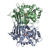
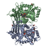
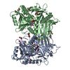
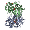
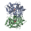
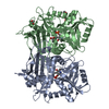
| Title | HUMAN BRANCHED CHAIN AMINO ACID AMINOTRANSFERASE : THREE DIMENSIONAL STRUCTURE OF THE ENZYME IN ITS PYRIDOXAMINE PHOSPHATE FORM. | ||||||
 Components Components | BRANCHED-CHAIN AMINO ACID AMINOTRANSFERASE, MITOCHONDRIAL | ||||||
 Keywords Keywords | TRANSFERASE / FOLD TYPE IV | ||||||
| Function / homology |  Function and homology information Function and homology informationregulation of hormone levels / branched-chain-amino-acid:2-oxoglutarate transaminase activity / L-valine:2-oxoglutarate transaminase activity / L-isoleucine:2-oxoglutarate transaminase activity / L-leucine:2-oxoglutarate transaminase activity / L-isoleucine catabolic process / branched-chain amino acid biosynthetic process / branched-chain-amino-acid transaminase / L-leucine biosynthetic process / branched-chain amino acid catabolic process ...regulation of hormone levels / branched-chain-amino-acid:2-oxoglutarate transaminase activity / L-valine:2-oxoglutarate transaminase activity / L-isoleucine:2-oxoglutarate transaminase activity / L-leucine:2-oxoglutarate transaminase activity / L-isoleucine catabolic process / branched-chain amino acid biosynthetic process / branched-chain-amino-acid transaminase / L-leucine biosynthetic process / branched-chain amino acid catabolic process / Branched-chain amino acid catabolism / L-valine biosynthetic process / cellular response to leukemia inhibitory factor / brown fat cell differentiation / lipid metabolic process / mitochondrial matrix / mitochondrion / nucleoplasm Similarity search - Function | ||||||
| Biological species |  Homo sapiens (human) Homo sapiens (human) | ||||||
| Method |  X-RAY DIFFRACTION / SYNCHROTRON / MOLECULAR REPLACEMENT / Resolution: 1.9 Å X-RAY DIFFRACTION / SYNCHROTRON / MOLECULAR REPLACEMENT / Resolution: 1.9 Å | ||||||
 Authors Authors | Yennawar, N.H. / Conway, M.E. / Yennawar, H.P. / Farber, G.K. / Hutson, S.M. | ||||||
 Citation Citation | Journal: Biochemistry / Year: 2002 Title: Crystal structures of human mitochondrial branched chain aminotransferase reaction intermediates: ketimine and pyridoxamine phosphate forms Authors: Yennawar, N.H. / Conway, M.E. / Yennawar, H.P. / Farber, G.K. / Hutson, S.M. #1: Journal: Acta Crystallogr.,Sect.D / Year: 2001Title: The Structure of Human Mitochondrial Branched-Chain Aminotransferase Authors: Yennawar, N. / Dunbar, J. / Conway, M. / Hutson, S. / Farber, G. #2: Journal: Biochim.Biophys.Acta / Year: 1997Title: Cloning of the Rat and Human Mitochondrial Branched Amino Acid Aminotransferase (Bcatm). Authors: Bledsoe, R.K. / Dawson, P.A. / Hutson, S.M. | ||||||
| History |
|
- Structure visualization
| Structure viewer | Molecule: MolmilJmol/JSmol |
|---|
- Downloads & links
-Download
| PDBx/mmCIF format | 1kta.cif.gz | 161.9 KB | Display | PDBx/mmCIF format |
|---|---|---|---|---|
| PDB format | pdb1kta.ent.gz | 127.7 KB | Display | PDB format |
| PDBx/mmJSON format | 1kta.json.gz | Tree view | PDBx/mmJSON format | |
| Others |  Other downloads Other downloads |
-Validation report
| Arichive directory | https://data.pdbj.org/pub/pdb/validation_reports/kt/1ktaftp://data.pdbj.org/pub/pdb/validation_reports/kt/1kta | HTTPS FTP |
|---|
-Related structure data


-Links
 PDBj
PDBj


- Assembly
| Deposited unit | 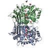
| ||||||||
|---|---|---|---|---|---|---|---|---|---|
| 1 |
| ||||||||
| Unit cell |
|
-Components
-Protein , 1 types, 2 molecules AB
| #1: Protein | Mass: 41368.016 Da / Num. of mol.: 2 Source method: isolated from a genetically manipulated source Source: (gene. exp.) Homo sapiens (human) / Plasmid: PET-28A / Species (production host): Escherichia coli / Production host:  References: UniProt: O15382, branched-chain-amino-acid transaminase |
|---|
-Non-polymers , 5 types, 254 molecules 


| #2: Chemical |  Mass: 248.173 Da / Num. of mol.: 2 / Source method: obtained synthetically / Formula: C8H13N2O5P Mass: 248.173 Da / Num. of mol.: 2 / Source method: obtained synthetically / Formula: C8H13N2O5P#3: Chemical | ChemComp-ACY /  Mass: 60.052 Da / Num. of mol.: 7 / Source method: obtained synthetically / Formula: C2H4O2 Mass: 60.052 Da / Num. of mol.: 7 / Source method: obtained synthetically / Formula: C2H4O2#4: Chemical | ChemComp-KIV / |  Mass: 116.115 Da / Num. of mol.: 1 / Source method: obtained synthetically / Formula: C5H8O3 Mass: 116.115 Da / Num. of mol.: 1 / Source method: obtained synthetically / Formula: C5H8O3#5: Chemical | ChemComp-GOL / |  Mass: 92.094 Da / Num. of mol.: 1 / Source method: obtained synthetically / Formula: C3H8O3 Mass: 92.094 Da / Num. of mol.: 1 / Source method: obtained synthetically / Formula: C3H8O3#6: Water | ChemComp-HOH / | Mass: 18.015 Da / Num. of mol.: 243 / Source method: isolated from a natural source / Formula: H2O |
|---|
-Experimental details
-Experiment
| Experiment | Method: X-RAY DIFFRACTION / Number of used crystals: 1 |
|---|
- Sample preparation
| Crystal | Density Matthews: 2.38 Å3/Da / Density % sol: 48.35 % | ||||||||||||||||||||||||||||||||||||||||
|---|---|---|---|---|---|---|---|---|---|---|---|---|---|---|---|---|---|---|---|---|---|---|---|---|---|---|---|---|---|---|---|---|---|---|---|---|---|---|---|---|---|
| Crystal grow | pH: 6.9 / Details: pH 6.90 | ||||||||||||||||||||||||||||||||||||||||
| Crystal grow | *PLUS pH: 7 / Method: vapor diffusion, hanging dropDetails: Yennawar, N., (2001) Acta Crystallogr., Sect.D, 57, 506. | ||||||||||||||||||||||||||||||||||||||||
| Components of the solutions | *PLUS
|
-Data collection
| Diffraction | Mean temperature: 223 K |
|---|---|
| Diffraction source | Source: SYNCHROTRON / Site: ALS  / Beamline: 5.0.2 / Wavelength: 1.0721 / Beamline: 5.0.2 / Wavelength: 1.0721 |
| Detector | Detector: CCD / Date: Apr 20, 1999 |
| Radiation | Monochromator: SI(111) DOUBLE CRYSTAL / Protocol: SINGLE WAVELENGTH / Monochromatic (M) / Laue (L): M / Scattering type: x-ray |
| Radiation wavelength | Wavelength: 1.0721 Å / Relative weight: 1 |
| Reflection | Resolution: 1.9→20 Å / Num. obs: 62273 / % possible obs: 98.4 % / Observed criterion σ(I): 0 / Redundancy: 1.6 % / Biso Wilson estimate: 27 Å2 / Rmerge(I) obs: 0.057 / Net I/σ(I): 0.215 |
| Reflection shell | Resolution: 1.9→1.93 Å / Redundancy: 1.6 % / Rmerge(I) obs: 0.403 / Mean I/σ(I) obs: 0.022 / % possible all: 91.1 |
| Reflection | *PLUS Highest resolution: 1.9 Å / Lowest resolution: 20 Å / Num. measured all: 246448 |
| Reflection shell | *PLUS % possible obs: 91.1 % / Mean I/σ(I) obs: 2.2 |
- Processing
| Software |
| ||||||||||||||||||||||||||||||||||||||||||||||||||||||||||||||||||||||||||||||||
|---|---|---|---|---|---|---|---|---|---|---|---|---|---|---|---|---|---|---|---|---|---|---|---|---|---|---|---|---|---|---|---|---|---|---|---|---|---|---|---|---|---|---|---|---|---|---|---|---|---|---|---|---|---|---|---|---|---|---|---|---|---|---|---|---|---|---|---|---|---|---|---|---|---|---|---|---|---|---|---|---|---|
| Refinement | Method to determine structure: MOLECULAR REPLACEMENT / Resolution: 1.9→19.96 Å / Rfactor Rfree error: 0.005 / Data cutoff high rms absF: 2153684.73 / Isotropic thermal model: RESTRAINED / Cross valid method: THROUGHOUT / σ(F): 2 / Stereochemistry target values: ENGH & HUBER Details: THE ATOMS OF THE HET GROUP KIV REPRESENT THE PRODUCT, KETO ISO VALERATE OF THE REACTION. THESE ATOMS APPEAR ONLY AT LOW CONTOUR LEVELS.
| ||||||||||||||||||||||||||||||||||||||||||||||||||||||||||||||||||||||||||||||||
| Solvent computation | Solvent model: FLAT MODEL / Bsol: 43.6841 Å2 / ksol: 0.333217 e/Å3 | ||||||||||||||||||||||||||||||||||||||||||||||||||||||||||||||||||||||||||||||||
| Displacement parameters | Biso mean: 36.5 Å2
| ||||||||||||||||||||||||||||||||||||||||||||||||||||||||||||||||||||||||||||||||
| Refine analyze |
| ||||||||||||||||||||||||||||||||||||||||||||||||||||||||||||||||||||||||||||||||
| Refinement step | Cycle: LAST / Resolution: 1.9→19.96 Å
| ||||||||||||||||||||||||||||||||||||||||||||||||||||||||||||||||||||||||||||||||
| Refine LS restraints |
| ||||||||||||||||||||||||||||||||||||||||||||||||||||||||||||||||||||||||||||||||
| Refine LS restraints NCS | NCS model details: CONSTRAINED | ||||||||||||||||||||||||||||||||||||||||||||||||||||||||||||||||||||||||||||||||
| LS refinement shell | Resolution: 1.9→2.02 Å / Rfactor Rfree error: 0.015 / Total num. of bins used: 6
| ||||||||||||||||||||||||||||||||||||||||||||||||||||||||||||||||||||||||||||||||
| Xplor file |
| ||||||||||||||||||||||||||||||||||||||||||||||||||||||||||||||||||||||||||||||||
| Refinement | *PLUS Highest resolution: 1.9 Å / Lowest resolution: 20 Å / Rfactor Rwork: 0.24 | ||||||||||||||||||||||||||||||||||||||||||||||||||||||||||||||||||||||||||||||||
| Solvent computation | *PLUS | ||||||||||||||||||||||||||||||||||||||||||||||||||||||||||||||||||||||||||||||||
| Displacement parameters | *PLUS | ||||||||||||||||||||||||||||||||||||||||||||||||||||||||||||||||||||||||||||||||
| Refine LS restraints | *PLUS
|
