Mass: 4546.990 Da / Num. of mol.: 1 / Source method: obtained synthetically
#3: Protein
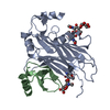
DEADRINGERPROTEIN
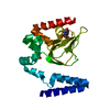
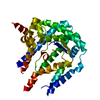
Mass: 16145.514 Da / Num. of mol.: 1 / Fragment: A/T Rich Interaction Domain / Mutation: F355L Source method: isolated from a genetically manipulated source Source: (gene. exp.) Drosophila melanogaster (fruit fly) / Gene: dri / Plasmid: pGEX-4T-1 / Species (production host): Escherichia coli / Production host: Escherichia coli BL21 (bacteria) / Strain (production host): BL21 / References: UniProt: Q24573
-
Experimental details
-
Experiment
Experiment
Method: SOLUTION NMR
NMR experiment
Conditions-ID
Experiment-ID
Solution-ID
Type
1
1
1
3D 13C-separated NOESY
1
2
1
3D 15N-separated NOESY
1
3
1
HNHA
1
4
2
3D 13C-separated NOESY
1
5
2
2D F1F2 13C-filtered NOESY
1
6
1
3D F1 13C15N-filtered F2 13C-edited NOESY
1
7
1
3D F1 13C15N-filtered F2 15N-edited NOESY
1
8
2
2D F2 13C-filtered NOESY
1
9
2
2D F1 13C-filtered JUNSY
1
10
1
3D HNHB
NMR details
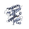
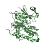
Text: The structure was determined using triple-resonance NMR methods along with various isotope-filtered techniques. Residues 1-2 and 134-139 are disordered in solution.
-
Sample preparation
Details
Solution-ID
Contents
Solvent system
1
2.0mM Dead ringer-DNA complex (protein U-13C,15N, DNA NA) 20mM Tris U-2H, 0.01% NaN3 NA, 0.5mM EDTA NA, 5mM DTT U-2H
93% H2O/7% D2O
2
1.4mM Dead ringer-DNA complex (protein U-13C,15N, DNA NA) 20mM Tris U-2H, 0.01% NaN3 NA, 0.5mM EDTA NA, 5mM DTT U-2H
In the structure databanks used in Yorodumi, some data are registered as the other names, "COVID-19 virus" and "2019-nCoV". Here are the details of the virus and the list of structure data.
Jan 31, 2019. EMDB accession codes are about to change! (news from PDBe EMDB page)
EMDB accession codes are about to change! (news from PDBe EMDB page)
The allocation of 4 digits for EMDB accession codes will soon come to an end. Whilst these codes will remain in use, new EMDB accession codes will include an additional digit and will expand incrementally as the available range of codes is exhausted. The current 4-digit format prefixed with “EMD-” (i.e. EMD-XXXX) will advance to a 5-digit format (i.e. EMD-XXXXX), and so on. It is currently estimated that the 4-digit codes will be depleted around Spring 2019, at which point the 5-digit format will come into force.
The EM Navigator/Yorodumi systems omit the EMD- prefix.
Related info.:Q: What is EMD? / ID/Accession-code notation in Yorodumi/EM Navigator
Yorodumi is a browser for structure data from EMDB, PDB, SASBDB, etc.
This page is also the successor to EM Navigator detail page, and also detail information page/front-end page for Omokage search.
The word "yorodu" (or yorozu) is an old Japanese word meaning "ten thousand". "mi" (miru) is to see.
Related info.:EMDB / PDB / SASBDB / Comparison of 3 databanks / Yorodumi Search / Aug 31, 2016. New EM Navigator & Yorodumi / Yorodumi Papers / Jmol/JSmol / Function and homology information / Changes in new EM Navigator and Yorodumi
 Movie
Movie Controller


 Open data
 Basic information
 Components
 Keywords
 Function and homology information
 Authors
 Citation
 Structure visualization
 Downloads & links
 Other downloads


 PDBj
PDBj


 Assembly
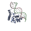

 Sample preparation
 Processing
 NMRPipe