 Movie
Movie Controller


+ Open data

- Basic information
| Entry | Database: PDB / ID: 4zk9 | |||||||||
|---|---|---|---|---|---|---|---|---|---|---|
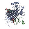
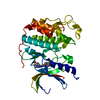
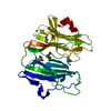
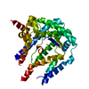
| Title | The chemokine binding protein of orf virus complexed with CCL2 | |||||||||
 Components Components |
| |||||||||
 Keywords Keywords | Viral Protein/cytokine / Complex / Orf virus chemokine binding protein / Human C-C motif chemokine 2 / Viral Protein-cytokine complex | |||||||||
| Function / homology |  Function and homology information Function and homology informationhelper T cell extravasation / chemokine (C-C motif) ligand 2 signaling pathway / CCR2 chemokine receptor binding / negative regulation of natural killer cell chemotaxis / astrocyte cell migration / chemokine receptor binding / ATF4 activates genes in response to endoplasmic reticulum stress / CCR chemokine receptor binding / negative regulation of glial cell apoptotic process / positive regulation of apoptotic cell clearance ...helper T cell extravasation / chemokine (C-C motif) ligand 2 signaling pathway / CCR2 chemokine receptor binding / negative regulation of natural killer cell chemotaxis / astrocyte cell migration / chemokine receptor binding / ATF4 activates genes in response to endoplasmic reticulum stress / CCR chemokine receptor binding / negative regulation of glial cell apoptotic process / positive regulation of apoptotic cell clearance / NFE2L2 regulating inflammation associated genes / cellular homeostasis / eosinophil chemotaxis / positive regulation of glutamate receptor signaling pathway / negative regulation of vascular endothelial cell proliferation / chemokine activity / Chemokine receptors bind chemokines / negative regulation of G1/S transition of mitotic cell cycle / positive regulation of macrophage chemotaxis / chemoattractant activity / positive regulation of calcium ion import / Interleukin-10 signaling / humoral immune response / macrophage chemotaxis / monocyte chemotaxis / positive regulation of endothelial cell apoptotic process / cellular response to interleukin-1 / G protein-coupled receptor signaling pathway, coupled to cyclic nucleotide second messenger / cell surface receptor signaling pathway via JAK-STAT / positive regulation of synaptic transmission, glutamatergic / cytoskeleton organization / cellular response to fibroblast growth factor stimulus / response to bacterium / sensory perception of pain / viral genome replication / animal organ morphogenesis / chemokine-mediated signaling pathway / cellular response to type II interferon / cellular response to tumor necrosis factor / chemotaxis / positive regulation of T cell activation / cytokine-mediated signaling pathway / regulation of cell shape / antimicrobial humoral immune response mediated by antimicrobial peptide / positive regulation of cytosolic calcium ion concentration / cellular response to lipopolysaccharide / angiogenesis / Interleukin-4 and Interleukin-13 signaling / negative regulation of neuron apoptotic process / protein phosphorylation / protein kinase activity / cell surface receptor signaling pathway / cell adhesion / positive regulation of cell migration / inflammatory response / G protein-coupled receptor signaling pathway / signaling receptor binding / positive regulation of gene expression / signal transduction / : / extracellular region / membrane / identical protein binding Similarity search - Function | |||||||||
| Biological species |  Orf virus Orf virus Homo sapiens (human) Homo sapiens (human) | |||||||||
| Method |  X-RAY DIFFRACTION / SYNCHROTRON / MOLECULAR REPLACEMENT / Resolution: 2.6 Å X-RAY DIFFRACTION / SYNCHROTRON / MOLECULAR REPLACEMENT / Resolution: 2.6 Å | |||||||||
 Authors Authors | Knapp, K.M. / Nakatani, Y. / Krause, K.L. | |||||||||
 Citation Citation | Journal: Structure / Year: 2015 Title: Structures of Orf Virus Chemokine Binding Protein in Complex with Host Chemokines Reveal Clues to Broad Binding Specificity. Authors: Counago, R.M. / Knapp, K.M. / Nakatani, Y. / Fleming, S.B. / Corbett, M. / Wise, L.M. / Mercer, A.A. / Krause, K.L. | |||||||||
| History |
|
- Structure visualization
| Structure viewer | Molecule: MolmilJmol/JSmol |
|---|
- Downloads & links
-Download
| PDBx/mmCIF format | 4zk9.cif.gz | 122.2 KB | Display | PDBx/mmCIF format |
|---|---|---|---|---|
| PDB format | pdb4zk9.ent.gz | 92.8 KB | Display | PDB format |
| PDBx/mmJSON format | 4zk9.json.gz | Tree view | PDBx/mmJSON format | |
| Others |  Other downloads Other downloads |
-Validation report
| Arichive directory | https://data.pdbj.org/pub/pdb/validation_reports/zk/4zk9ftp://data.pdbj.org/pub/pdb/validation_reports/zk/4zk9 | HTTPS FTP |
|---|
-Related structure data
| Related structure data |  4p5iSC  4zkbC 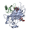 4zkcC C: citing same article ( S: Starting model for refinement |
|---|---|
| Similar structure data |


-Links
 PDBj
PDBj


- Assembly
| Deposited unit | 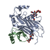
| ||||||||
|---|---|---|---|---|---|---|---|---|---|
| 1 |
| ||||||||
| Unit cell |
|
-Components
| #1: Protein | Mass: 30374.613 Da / Num. of mol.: 1 / Fragment: UNP residues 17-286 Source method: isolated from a genetically manipulated source Source: (gene. exp.) Orf virus (strain NZ2) / Plasmid: PTT5 / Cell line (production host): HEK 293-6E / Production host: Homo sapiens (human) / References: UniProt: Q2F862 | ||
|---|---|---|---|
| #2: Protein | Mass: 9666.073 Da / Num. of mol.: 1 / Fragment: UNP residues 17-99 Source method: isolated from a genetically manipulated source Source: (gene. exp.) Homo sapiens (human) / Gene: CCL2, MCP1, SCYA2 / Plasmid: pTT5 / Cell line (production host): HEK 293-6E / Production host: Homo sapiens (human) / References: UniProt: P13500 | ||
| #3: Polysaccharide | Source method: isolated from a genetically manipulated source Has protein modification | Y | |
-Experimental details
-Experiment
| Experiment | Method: X-RAY DIFFRACTION / Number of used crystals: 1 |
|---|
- Sample preparation
| Crystal | Density Matthews: 3.63 Å3/Da / Density % sol: 66.16 % |
|---|---|
| Crystal grow | Temperature: 289 K / Method: vapor diffusion, hanging drop / pH: 5.6 Details: 0.2M potassium sodium tartrate tetrahydrate, 0.1M sodium citrate tribasic dihydrate pH5.6, 2.0M ammonium citrate |
-Data collection
| Diffraction | Mean temperature: 93.2 K |
|---|---|
| Diffraction source | Source: SYNCHROTRON / Site: Australian Synchrotron  / Beamline: MX1 / Wavelength: 0.95369 Å / Beamline: MX1 / Wavelength: 0.95369 Å |
| Detector | Type: ADSC QUANTUM 210r / Detector: CCD / Date: Mar 10, 2011 |
| Radiation | Protocol: SINGLE WAVELENGTH / Monochromatic (M) / Laue (L): M / Scattering type: x-ray |
| Radiation wavelength | Wavelength: 0.95369 Å / Relative weight: 1 |
| Reflection | Resolution: 2.6→38.54 Å / Num. obs: 18835 / % possible obs: 100 % / Redundancy: 14.2 % / Biso Wilson estimate: 62.6 Å2 / Rmerge(I) obs: 0.067 / Net I/σ(I): 31.9 |
| Reflection shell | Resolution: 2.6→2.74 Å / Redundancy: 14.6 % / Rmerge(I) obs: 0.893 / Mean I/σ(I) obs: 3.1 / % possible all: 100 |
- Processing
| Software |
| ||||||||||||||||||||||||||||||||||||||||||||||||||||||||||||||||||||||||||||||||||||||||||||||||||||||||||||||||||||||||||||||||||||||||||||||||||||||||||||||||||||||||||||||||||||||
|---|---|---|---|---|---|---|---|---|---|---|---|---|---|---|---|---|---|---|---|---|---|---|---|---|---|---|---|---|---|---|---|---|---|---|---|---|---|---|---|---|---|---|---|---|---|---|---|---|---|---|---|---|---|---|---|---|---|---|---|---|---|---|---|---|---|---|---|---|---|---|---|---|---|---|---|---|---|---|---|---|---|---|---|---|---|---|---|---|---|---|---|---|---|---|---|---|---|---|---|---|---|---|---|---|---|---|---|---|---|---|---|---|---|---|---|---|---|---|---|---|---|---|---|---|---|---|---|---|---|---|---|---|---|---|---|---|---|---|---|---|---|---|---|---|---|---|---|---|---|---|---|---|---|---|---|---|---|---|---|---|---|---|---|---|---|---|---|---|---|---|---|---|---|---|---|---|---|---|---|---|---|---|---|
| Refinement | Method to determine structure: MOLECULAR REPLACEMENT Starting model: 4P5I Resolution: 2.6→38.54 Å / Cor.coef. Fo:Fc: 0.93 / Cor.coef. Fo:Fc free: 0.901 / SU B: 19.925 / SU ML: 0.198 / Cross valid method: THROUGHOUT / ESU R: 0.294 / ESU R Free: 0.254 / Stereochemistry target values: MAXIMUM LIKELIHOOD / Details: HYDROGENS HAVE BEEN ADDED IN THE RIDING POSITIONS
| ||||||||||||||||||||||||||||||||||||||||||||||||||||||||||||||||||||||||||||||||||||||||||||||||||||||||||||||||||||||||||||||||||||||||||||||||||||||||||||||||||||||||||||||||||||||
| Solvent computation | Ion probe radii: 0.8 Å / Shrinkage radii: 0.8 Å / VDW probe radii: 1.2 Å / Solvent model: MASK | ||||||||||||||||||||||||||||||||||||||||||||||||||||||||||||||||||||||||||||||||||||||||||||||||||||||||||||||||||||||||||||||||||||||||||||||||||||||||||||||||||||||||||||||||||||||
| Displacement parameters | Biso mean: 77.474 Å2
| ||||||||||||||||||||||||||||||||||||||||||||||||||||||||||||||||||||||||||||||||||||||||||||||||||||||||||||||||||||||||||||||||||||||||||||||||||||||||||||||||||||||||||||||||||||||
| Refinement step | Cycle: LAST / Resolution: 2.6→38.54 Å
| ||||||||||||||||||||||||||||||||||||||||||||||||||||||||||||||||||||||||||||||||||||||||||||||||||||||||||||||||||||||||||||||||||||||||||||||||||||||||||||||||||||||||||||||||||||||
| Refine LS restraints |
|