 Movie
Movie Controller

[English] 日本語
 Yorodumi
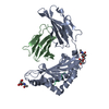
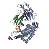
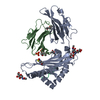
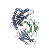
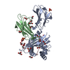
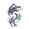
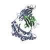
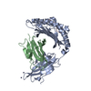
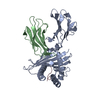
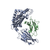
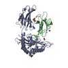
- PDB-1kpu: High resolution crystal structure of the MHC class I complex H-2K... -
+ Open data

- Basic information
| Entry | Database: PDB / ID: 1kpu | |||||||||
|---|---|---|---|---|---|---|---|---|---|---|
| Title | High resolution crystal structure of the MHC class I complex H-2Kb/VSV8 | |||||||||
 Components Components |
| |||||||||
 Keywords Keywords | IMMUNE SYSTEM / MAJOR HISTOCOMPATIBILITY COMPLEX / PEPTIDE-MHC | |||||||||
| Function / homology |  Function and homology information Function and homology informationEndosomal/Vacuolar pathway / DAP12 interactions / Antigen Presentation: Folding, assembly and peptide loading of class I MHC / ER-Phagosome pathway / DAP12 signaling / Immunoregulatory interactions between a Lymphoid and a non-Lymphoid cell / helical viral capsid / antigen processing and presentation of exogenous peptide antigen via MHC class I / inner ear development / antigen processing and presentation of endogenous peptide antigen via MHC class I via ER pathway, TAP-dependent ...Endosomal/Vacuolar pathway / DAP12 interactions / Antigen Presentation: Folding, assembly and peptide loading of class I MHC / ER-Phagosome pathway / DAP12 signaling / Immunoregulatory interactions between a Lymphoid and a non-Lymphoid cell / helical viral capsid / antigen processing and presentation of exogenous peptide antigen via MHC class I / inner ear development / antigen processing and presentation of endogenous peptide antigen via MHC class I via ER pathway, TAP-dependent / cellular defense response / Neutrophil degranulation / lumenal side of endoplasmic reticulum membrane / cellular response to iron(III) ion / negative regulation of forebrain neuron differentiation / antigen processing and presentation of exogenous protein antigen via MHC class Ib, TAP-dependent / iron ion transport / peptide antigen assembly with MHC class I protein complex / regulation of iron ion transport / regulation of erythrocyte differentiation / HFE-transferrin receptor complex / response to molecule of bacterial origin / MHC class I peptide loading complex / T cell mediated cytotoxicity / positive regulation of T cell cytokine production / antigen processing and presentation of endogenous peptide antigen via MHC class I / MHC class I protein complex / peptide antigen binding / positive regulation of receptor-mediated endocytosis / negative regulation of neurogenesis / cellular response to nicotine / positive regulation of T cell mediated cytotoxicity / multicellular organismal-level iron ion homeostasis / phagocytic vesicle membrane / negative regulation of epithelial cell proliferation / sensory perception of smell / positive regulation of cellular senescence / T cell differentiation in thymus / negative regulation of neuron projection development / protein refolding / viral nucleocapsid / protein homotetramerization / amyloid fibril formation / intracellular iron ion homeostasis / host cell cytoplasm / learning or memory / defense response to bacterium / ribonucleoprotein complex / external side of plasma membrane / structural molecule activity / Golgi apparatus / protein homodimerization activity / extracellular space / RNA binding / cytosol Similarity search - Function | |||||||||
| Biological species |  | |||||||||
| Method |  X-RAY DIFFRACTION / SYNCHROTRON / MOLECULAR REPLACEMENT / Resolution: 1.5 Å X-RAY DIFFRACTION / SYNCHROTRON / MOLECULAR REPLACEMENT / Resolution: 1.5 Å | |||||||||
 Authors Authors | Rudolph, M.G. / Wilson, I.A. | |||||||||
 Citation Citation | Journal: To be Published Title: High Resolution Crystal Structure of H-2Kb/VSV8 Authors: Rudolph, M.G. / Wilson, I.A. #1: Journal: Science / Year: 1992Title: Crystal Structures of Two Viral Peptides in Complex with Murine MHC class I H-2Kb Authors: Fremont, D.H. / Matsumura, M. / Stura, E.A. / Peterson, P.A. / Wilson, I.A. | |||||||||
| History |
|
- Structure visualization
| Structure viewer | Molecule: MolmilJmol/JSmol |
|---|
- Downloads & links
-Download
| PDBx/mmCIF format | 1kpu.cif.gz | 104.5 KB | Display | PDBx/mmCIF format |
|---|---|---|---|---|
| PDB format | pdb1kpu.ent.gz | 78.8 KB | Display | PDB format |
| PDBx/mmJSON format | 1kpu.json.gz | Tree view | PDBx/mmJSON format | |
| Others |  Other downloads Other downloads |
-Validation report
| Summary document | 1kpu_validation.pdf.gz | 864.9 KB | Display | wwPDB validaton report |
|---|---|---|---|---|
| Full document | 1kpu_full_validation.pdf.gz | 869.9 KB | Display | |
| Data in XML | 1kpu_validation.xml.gz | 21.7 KB | Display | |
| Data in CIF | 1kpu_validation.cif.gz | 32.4 KB | Display | |
| Arichive directory | https://data.pdbj.org/pub/pdb/validation_reports/kp/1kpuftp://data.pdbj.org/pub/pdb/validation_reports/kp/1kpu | HTTPS FTP |
-Related structure data
| Related structure data | 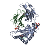 1kpvC  2vaaS S: Starting model for refinement C: citing same article ( |
|---|---|
| Similar structure data |


-Links
 PDBj
PDBj


- Assembly
| Deposited unit | 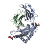
| ||||||||
|---|---|---|---|---|---|---|---|---|---|
| 1 |
| ||||||||
| Unit cell |
|
-Components
-Protein , 2 types, 2 molecules AB
| #1: Protein | Mass: 31648.322 Da / Num. of mol.: 1 Fragment: extracellular domain, sequence database residues 22-295, numbered 1-274 Source method: isolated from a genetically manipulated source Source: (gene. exp.)  |
|---|---|
| #2: Protein | Mass: 11704.359 Da / Num. of mol.: 1 / Fragment: sequence database residues 21-119, numbered 1-99 Source method: isolated from a genetically manipulated source Source: (gene. exp.) |
-Protein/peptide , 1 types, 1 molecules P
| #3: Protein/peptide | Mass: 956.078 Da / Num. of mol.: 1 / Fragment: sequence database residues 52-59, numbered 1-8 / Source method: obtained synthetically Details: THE PEPTIDE WAS CHEMICALLY SYNTHESIZED. THE SEQUENCE OF THE PEPTIDE IS FOUND NATURALLY IN VESICULAR STOMATITIS VIRUS. References: GenBank: 534831, UniProt: Q88992*PLUS |
|---|
-Sugars , 2 types, 2 molecules 
| #4: Polysaccharide | 2-acetamido-2-deoxy-beta-D-glucopyranose-(1-4)-[alpha-L-fucopyranose-(1-6)]2-acetamido-2-deoxy-beta- ...2-acetamido-2-deoxy-beta-D-glucopyranose-(1-4)-[alpha-L-fucopyranose-(1-6)]2-acetamido-2-deoxy-beta-D-glucopyranose Source method: isolated from a genetically manipulated source |
|---|---|
| #5: Sugar | ChemComp-NAG /  Type: D-saccharide, beta linking / Mass: 221.208 Da / Num. of mol.: 1 Type: D-saccharide, beta linking / Mass: 221.208 Da / Num. of mol.: 1Source method: isolated from a genetically manipulated source Formula: C8H15NO6 |
-Non-polymers , 2 types, 418 molecules 

| #6: Chemical |  Mass: 118.174 Da / Num. of mol.: 2 / Source method: obtained synthetically / Formula: C6H14O2 / Comment: precipitant*YM Mass: 118.174 Da / Num. of mol.: 2 / Source method: obtained synthetically / Formula: C6H14O2 / Comment: precipitant*YM#7: Water | ChemComp-HOH / | Mass: 18.015 Da / Num. of mol.: 416 / Source method: isolated from a natural source / Formula: H2O |
|---|
-Details
| Has protein modification | Y |
|---|
-Experimental details
-Experiment
| Experiment | Method: X-RAY DIFFRACTION / Number of used crystals: 1 |
|---|
- Sample preparation
| Crystal | Density Matthews: 3.06 Å3/Da / Density % sol: 59.8 % |
|---|---|
| Crystal grow | Temperature: 295 K / Method: vapor diffusion, sitting drop / pH: 6.5 Details: K/NA PHOSPHATE, MPD, pH 6.5, VAPOR DIFFUSION, SITTING DROP, temperature 295K |
-Data collection
| Diffraction | Mean temperature: 100 K |
|---|---|
| Diffraction source | Source: SYNCHROTRON / Site: APS  / Beamline: 19-ID / Wavelength: 1.033 Å / Beamline: 19-ID / Wavelength: 1.033 Å |
| Detector | Type: CUSTOM-MADE / Detector: CCD / Date: May 26, 2001 |
| Radiation | Protocol: SINGLE WAVELENGTH / Monochromatic (M) / Laue (L): M / Scattering type: x-ray |
| Radiation wavelength | Wavelength: 1.033 Å / Relative weight: 1 |
| Reflection | Resolution: 1.5→42 Å / Num. obs: 81282 / % possible obs: 92.5 % / Observed criterion σ(F): 0 / Observed criterion σ(I): -3 / Redundancy: 6.3 % / Rsym value: 0.113 / Net I/σ(I): 9.8 |
| Reflection shell | Resolution: 1.5→1.55 Å / Redundancy: 3.7 % / Mean I/σ(I) obs: 2.9 / Num. unique all: 6180 / Rsym value: 0.431 / % possible all: 71.5 |
- Processing
| Software |
| ||||||||||||||||||||||||||||||||||||||||||||||||||||||||||||||||||||||||||||||||||||||||||||||||||||||||||||||||||||||||||||||||||
|---|---|---|---|---|---|---|---|---|---|---|---|---|---|---|---|---|---|---|---|---|---|---|---|---|---|---|---|---|---|---|---|---|---|---|---|---|---|---|---|---|---|---|---|---|---|---|---|---|---|---|---|---|---|---|---|---|---|---|---|---|---|---|---|---|---|---|---|---|---|---|---|---|---|---|---|---|---|---|---|---|---|---|---|---|---|---|---|---|---|---|---|---|---|---|---|---|---|---|---|---|---|---|---|---|---|---|---|---|---|---|---|---|---|---|---|---|---|---|---|---|---|---|---|---|---|---|---|---|---|---|---|
| Refinement | Method to determine structure: MOLECULAR REPLACEMENT Starting model: PDB entry 2vaa, chains A and B Resolution: 1.5→41.89 Å / Cor.coef. Fo:Fc: 0.957 / Cor.coef. Fo:Fc free: 0.942 / SU B: 1.75 / SU ML: 0.065 / Cross valid method: THROUGHOUT / σ(F): 0 / ESU R: 0.07 / ESU R Free: 0.071 / Stereochemistry target values: Engh & Huber / Details: HYDROGENS HAVE BEEN ADDED IN THE RIDING POSITIONS
| ||||||||||||||||||||||||||||||||||||||||||||||||||||||||||||||||||||||||||||||||||||||||||||||||||||||||||||||||||||||||||||||||||
| Solvent computation | Ion probe radii: 0.8 Å / Shrinkage radii: 0.8 Å / VDW probe radii: 1.4 Å / Solvent model: BABINET MODEL WITH MASK | ||||||||||||||||||||||||||||||||||||||||||||||||||||||||||||||||||||||||||||||||||||||||||||||||||||||||||||||||||||||||||||||||||
| Displacement parameters | Biso mean: 17.187 Å2
| ||||||||||||||||||||||||||||||||||||||||||||||||||||||||||||||||||||||||||||||||||||||||||||||||||||||||||||||||||||||||||||||||||
| Refinement step | Cycle: LAST / Resolution: 1.5→41.89 Å
| ||||||||||||||||||||||||||||||||||||||||||||||||||||||||||||||||||||||||||||||||||||||||||||||||||||||||||||||||||||||||||||||||||
| Refine LS restraints |
| ||||||||||||||||||||||||||||||||||||||||||||||||||||||||||||||||||||||||||||||||||||||||||||||||||||||||||||||||||||||||||||||||||
| LS refinement shell | Resolution: 1.5→1.54 Å / Total num. of bins used: 20
|
