 Movie
Movie Controller

[English] 日本語
 Yorodumi
- PDB-1kkt: Structure of P. citrinum alpha 1,2-mannosidase reveals the basis ... -
+ Open data

- Basic information
| Entry | Database: PDB / ID: 1kkt | |||||||||
|---|---|---|---|---|---|---|---|---|---|---|
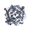
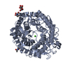
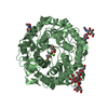
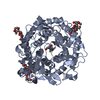
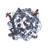
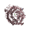
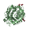
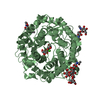
| Title | Structure of P. citrinum alpha 1,2-mannosidase reveals the basis for differences in specificity of the ER and Golgi Class I enzymes | |||||||||
 Components Components | Mannosyl-oligosaccharide alpha-1,2-mannosidase | |||||||||
 Keywords Keywords | HYDROLASE / (alpha/alpha)7-barrel | |||||||||
| Function / homology |  Function and homology information Function and homology informationmannosyl-oligosaccharide 1,2-alpha-mannosidase / mannosyl-oligosaccharide 1,2-alpha-mannosidase activity / ERAD pathway / carbohydrate metabolic process / calcium ion binding / endoplasmic reticulum / extracellular region / membrane Similarity search - Function | |||||||||
| Biological species |  Penicillium citrinum (fungus) Penicillium citrinum (fungus) | |||||||||
| Method |  X-RAY DIFFRACTION / MOLECULAR REPLACEMENT / Resolution: 2.2 Å X-RAY DIFFRACTION / MOLECULAR REPLACEMENT / Resolution: 2.2 Å | |||||||||
 Authors Authors | Lobsanov, Y.D. / Vallee, F. / Imberty, A. / Yoshida, T. / Yip, P. / Herscovics, A. / Howell, P.L. | |||||||||
 Citation Citation | Journal: J.Biol.Chem. / Year: 2002 Title: Structure of Penicillium citrinum alpha 1,2-mannosidase reveals the basis for differences in specificity of the endoplasmic reticulum and Golgi class I enzymes. Authors: Lobsanov, Y.D. / Vallee, F. / Imberty, A. / Yoshida, T. / Yip, P. / Herscovics, A. / Howell, P.L. | |||||||||
| History |
|
- Structure visualization
| Structure viewer | Molecule: MolmilJmol/JSmol |
|---|
- Downloads & links
-Download
| PDBx/mmCIF format | 1kkt.cif.gz | 203.1 KB | Display | PDBx/mmCIF format |
|---|---|---|---|---|
| PDB format | pdb1kkt.ent.gz | 163.8 KB | Display | PDB format |
| PDBx/mmJSON format | 1kkt.json.gz | Tree view | PDBx/mmJSON format | |
| Others |  Other downloads Other downloads |
-Validation report
| Arichive directory | https://data.pdbj.org/pub/pdb/validation_reports/kk/1kktftp://data.pdbj.org/pub/pdb/validation_reports/kk/1kkt | HTTPS FTP |
|---|
-Related structure data
| Related structure data |  1kreC  1krfC 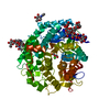 1dl2S  1fm1S S: Starting model for refinement C: citing same article ( |
|---|---|
| Similar structure data |


-Links
 PDBj
PDBj
- Assembly
| Deposited unit | 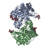
| ||||||||
|---|---|---|---|---|---|---|---|---|---|
| 1 | 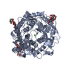
| ||||||||
| 2 | 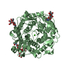
| ||||||||
| Unit cell |
| ||||||||
| Details | The biological assembly is a monomer |
-Components
| #1: Protein | Mass: 56626.570 Da / Num. of mol.: 2 Source method: isolated from a genetically manipulated source Source: (gene. exp.) Penicillium citrinum (fungus) / Gene: msdC / Plasmid: pTAPM1 / Production host: References: UniProt: P31723, mannosyl-oligosaccharide 1,2-alpha-mannosidase #2: Polysaccharide | 2-acetamido-2-deoxy-beta-D-glucopyranose-(1-4)-2-acetamido-2-deoxy-beta-D-glucopyranose Source method: isolated from a genetically manipulated source #3: Polysaccharide | Source method: isolated from a genetically manipulated source #4: Chemical |   Mass: 40.078 Da / Num. of mol.: 2 / Source method: obtained synthetically / Formula: Ca Mass: 40.078 Da / Num. of mol.: 2 / Source method: obtained synthetically / Formula: Ca#5: Water | ChemComp-HOH / |  Mass: 18.015 Da / Num. of mol.: 201 / Source method: isolated from a natural source / Formula: H2O Mass: 18.015 Da / Num. of mol.: 201 / Source method: isolated from a natural source / Formula: H2OHas protein modification | Y | |
|---|
-Experimental details
-Experiment
| Experiment | Method: X-RAY DIFFRACTION / Number of used crystals: 1 |
|---|
- Sample preparation
| Crystal | Density Matthews: 2.36 Å3/Da / Density % sol: 47.78 % | ||||||||||||||||||||||||||||||
|---|---|---|---|---|---|---|---|---|---|---|---|---|---|---|---|---|---|---|---|---|---|---|---|---|---|---|---|---|---|---|---|
| Crystal grow | Temperature: 293 K / Method: vapor diffusion, hanging drop / pH: 4.6 Details: PEG 6000, potassium phosphate, pH 4.6, VAPOR DIFFUSION, HANGING DROP, temperature 293K | ||||||||||||||||||||||||||||||
| Crystal grow | *PLUS pH: 5 | ||||||||||||||||||||||||||||||
| Components of the solutions | *PLUS
|
-Data collection
| Diffraction | Mean temperature: 293 K |
|---|---|
| Diffraction source | Source: ROTATING ANODE / Type: RIGAKU RU200 / Wavelength: 1.5418 Å |
| Detector | Type: MARRESEARCH / Detector: AREA DETECTOR / Date: Feb 26, 2000 / Details: mirrors |
| Radiation | Monochromator: YALE MIRRORS / Protocol: SINGLE WAVELENGTH / Monochromatic (M) / Laue (L): M / Scattering type: x-ray |
| Radiation wavelength | Wavelength: 1.5418 Å / Relative weight: 1 |
| Reflection | Resolution: 2.2→50 Å / Num. all: 49814 / Num. obs: 49814 / % possible obs: 92.6 % / Observed criterion σ(F): 0 / Observed criterion σ(I): 0 / Redundancy: 3.49 % / Rmerge(I) obs: 0.069 / Rsym value: 0.069 / Net I/σ(I): 16.3 |
| Reflection shell | Resolution: 2.2→2.25 Å / Num. unique all: 2151 / Rsym value: 0.242 / % possible all: 60.1 |
| Reflection | *PLUS Num. obs: 49836 / Num. measured all: 174227 |
| Reflection shell | *PLUS % possible obs: 60.1 % / Rmerge(I) obs: 0.243 |
- Processing
| Software |
| ||||||||||||||||||||||||||||||
|---|---|---|---|---|---|---|---|---|---|---|---|---|---|---|---|---|---|---|---|---|---|---|---|---|---|---|---|---|---|---|---|
| Refinement | Method to determine structure: MOLECULAR REPLACEMENT Starting model: PDB ENTRIES 1DL2 and 1FM1 Resolution: 2.2→50 Å / σ(F): 0 / Stereochemistry target values: Engh & Huber
| ||||||||||||||||||||||||||||||
| Displacement parameters |
| ||||||||||||||||||||||||||||||
| Refinement step | Cycle: LAST / Resolution: 2.2→50 Å
| ||||||||||||||||||||||||||||||
| Software | *PLUS Name: CNS / Version: 1 / Classification: refinement | ||||||||||||||||||||||||||||||
| Refinement | *PLUS Lowest resolution: 50 Å / σ(F): 0 / % reflection Rfree: 10 % / Rfactor obs: 0.193 | ||||||||||||||||||||||||||||||
| Solvent computation | *PLUS | ||||||||||||||||||||||||||||||
| Displacement parameters | *PLUS | ||||||||||||||||||||||||||||||
| Refine LS restraints | *PLUS
|
