 Movie
Movie Controller


+ Open data

- Basic information
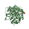
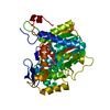
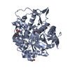
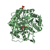
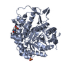
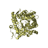
| Entry | Database: PDB / ID: 1hcu | ||||||
|---|---|---|---|---|---|---|---|
| Title | alpha-1,2-mannosidase from Trichoderma reesei | ||||||
 Components Components | ALPHA-1,2-MANNOSIDASE | ||||||
 Keywords Keywords | GLYCOSYLATION / GLYCOSYL HYDROLASE | ||||||
| Function / homology |  Function and homology information Function and homology informationmannosyl-oligosaccharide 1,2-alpha-mannosidase activity / Hydrolases; Glycosylases; Glycosidases, i.e. enzymes that hydrolyse O- and S-glycosyl compounds / ERAD pathway / carbohydrate metabolic process / calcium ion binding / endoplasmic reticulum / membrane Similarity search - Function | ||||||
| Biological species |  TRICHODERMA REESEI (fungus) TRICHODERMA REESEI (fungus) | ||||||
| Method |  X-RAY DIFFRACTION / SYNCHROTRON / MOLECULAR REPLACEMENT / Resolution: 2.37 Å X-RAY DIFFRACTION / SYNCHROTRON / MOLECULAR REPLACEMENT / Resolution: 2.37 Å | ||||||
 Authors Authors | Van Petegem, F. / Contreras, H. / Contreras, R. / Van Beeumen, J. | ||||||
 Citation Citation | Journal: J.Mol.Biol. / Year: 2001 Title: Trichoderma Reesei Alpha-1,2-Mannosidase: Structural Basis for the Cleavage of Four Consecutive Mannose Residues Authors: Van Petegem, F. / Contreras, H. / Contreras, R. / Van Beeumen, J. | ||||||
| History |
|
- Structure visualization
| Structure viewer | Molecule: MolmilJmol/JSmol |
|---|
- Downloads & links
-Download
| PDBx/mmCIF format | 1hcu.cif.gz | 393 KB | Display | PDBx/mmCIF format |
|---|---|---|---|---|
| PDB format | pdb1hcu.ent.gz | 321 KB | Display | PDB format |
| PDBx/mmJSON format | 1hcu.json.gz | Tree view | PDBx/mmJSON format | |
| Others |  Other downloads Other downloads |
-Validation report
| Arichive directory | https://data.pdbj.org/pub/pdb/validation_reports/hc/1hcuftp://data.pdbj.org/pub/pdb/validation_reports/hc/1hcu | HTTPS FTP |
|---|
-Related structure data
| Related structure data | 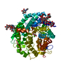 1dl2S S: Starting model for refinement |
|---|---|
| Similar structure data |


-Links
 PDBj
PDBj
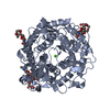
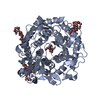
- Assembly
| Deposited unit | 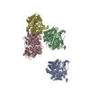
| ||||||||||||||||
|---|---|---|---|---|---|---|---|---|---|---|---|---|---|---|---|---|---|
| 1 | 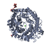
| ||||||||||||||||
| 2 | 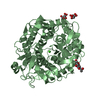
| ||||||||||||||||
| 3 | 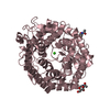
| ||||||||||||||||
| 4 | 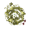
| ||||||||||||||||
| Unit cell |
| ||||||||||||||||
| Noncrystallographic symmetry (NCS) | NCS oper:
|
-Components
| #1: Protein | Mass: 54359.609 Da / Num. of mol.: 4 Source method: isolated from a genetically manipulated source Source: (gene. exp.) TRICHODERMA REESEI (fungus) / Production host: PICHIA PASTORIS (fungus)References: UniProt: Q9P8T8, mannosyl-oligosaccharide 1,2-alpha-mannosidase #2: Sugar | ChemComp-NAG /   Type: D-saccharide, beta linking / Mass: 221.208 Da / Num. of mol.: 10 Type: D-saccharide, beta linking / Mass: 221.208 Da / Num. of mol.: 10Source method: isolated from a genetically manipulated source Formula: C8H15NO6 #3: Chemical | ChemComp-CA /   Mass: 40.078 Da / Num. of mol.: 4 / Source method: obtained synthetically / Formula: Ca Mass: 40.078 Da / Num. of mol.: 4 / Source method: obtained synthetically / Formula: Ca#4: Water | ChemComp-HOH / |  Mass: 18.015 Da / Num. of mol.: 1094 / Source method: isolated from a natural source / Formula: H2O Mass: 18.015 Da / Num. of mol.: 1094 / Source method: isolated from a natural source / Formula: H2OHas protein modification | Y | |
|---|
-Experimental details
-Experiment
| Experiment | Method: X-RAY DIFFRACTION / Number of used crystals: 1 |
|---|
- Sample preparation
| Crystal | Density Matthews: 2.102 Å3/Da / Density % sol: 39.22 % | ||||||||||||||||||||||||||||||||||||||||||
|---|---|---|---|---|---|---|---|---|---|---|---|---|---|---|---|---|---|---|---|---|---|---|---|---|---|---|---|---|---|---|---|---|---|---|---|---|---|---|---|---|---|---|---|
| Crystal grow | Method: vapor diffusion, hanging drop / pH: 4 Details: 10MG/ML PROTEIN IN 0.1M TRIS-CL PH 7.5 MIX 2 MICROLITER OF PROTEIN SOLUTION WITH 2 MICROLITER OF WELL SOLUTION IN A HANGING DROP EXPERIMENT. THE WELL CONTAINS 0.1M NA-ACETATE PH 4.0, 12% PEG 35000, 0.3M CACL2 | ||||||||||||||||||||||||||||||||||||||||||
| Crystal grow | *PLUS pH: 7.4 / Method: vapor diffusion, hanging drop | ||||||||||||||||||||||||||||||||||||||||||
| Components of the solutions | *PLUS
|
-Data collection
| Diffraction | Mean temperature: 100 K |
|---|---|
| Diffraction source | Source: SYNCHROTRON / Site: EMBL/DESY, HAMBURG  / Beamline: X11 / Wavelength: 0.9 / Beamline: X11 / Wavelength: 0.9 |
| Detector | Type: MARRESEARCH / Detector: CCD / Date: Jan 15, 2000 |
| Radiation | Protocol: SINGLE WAVELENGTH / Monochromatic (M) / Laue (L): M / Scattering type: x-ray |
| Radiation wavelength | Wavelength: 0.9 Å / Relative weight: 1 |
| Reflection | Resolution: 2.37→20 Å / Num. obs: 73996 / % possible obs: 98.9 % / Redundancy: 4.94 % / Biso Wilson estimate: 20.4 Å2 / Rmerge(I) obs: 0.083 / Net I/σ(I): 20.01 |
| Reflection shell | Resolution: 2.37→2.45 Å / Redundancy: 4.14 % / Rmerge(I) obs: 0.214 / Mean I/σ(I) obs: 4.915 / % possible all: 95.1 |
| Reflection | *PLUS Num. measured all: 818429 |
| Reflection shell | *PLUS % possible obs: 95.1 % |
- Processing
| Software |
| ||||||||||||||||||||||||||||||||||||||||||||||||||||||||||||||||||||||||||||||||
|---|---|---|---|---|---|---|---|---|---|---|---|---|---|---|---|---|---|---|---|---|---|---|---|---|---|---|---|---|---|---|---|---|---|---|---|---|---|---|---|---|---|---|---|---|---|---|---|---|---|---|---|---|---|---|---|---|---|---|---|---|---|---|---|---|---|---|---|---|---|---|---|---|---|---|---|---|---|---|---|---|---|
| Refinement | Method to determine structure: MOLECULAR REPLACEMENT Starting model: PDB ENTRY 1DL2 Resolution: 2.37→20 Å / Rfactor Rfree error: 0.004 / Isotropic thermal model: RESTRAINED / Cross valid method: THROUGHOUT / σ(F): 0 Stereochemistry target values: MAXIMUM LIKELIHOOD USING AMPLITUDES
| ||||||||||||||||||||||||||||||||||||||||||||||||||||||||||||||||||||||||||||||||
| Solvent computation | Solvent model: FLAT MODEL / Bsol: 43.35 Å2 / ksol: 0.355 e/Å3 | ||||||||||||||||||||||||||||||||||||||||||||||||||||||||||||||||||||||||||||||||
| Displacement parameters | Biso mean: 21.6 Å2
| ||||||||||||||||||||||||||||||||||||||||||||||||||||||||||||||||||||||||||||||||
| Refine analyze |
| ||||||||||||||||||||||||||||||||||||||||||||||||||||||||||||||||||||||||||||||||
| Refinement step | Cycle: LAST / Resolution: 2.37→20 Å
| ||||||||||||||||||||||||||||||||||||||||||||||||||||||||||||||||||||||||||||||||
| Refine LS restraints |
| ||||||||||||||||||||||||||||||||||||||||||||||||||||||||||||||||||||||||||||||||
| LS refinement shell | Resolution: 2.37→2.52 Å / Rfactor Rfree error: 0.012 / Total num. of bins used: 6
| ||||||||||||||||||||||||||||||||||||||||||||||||||||||||||||||||||||||||||||||||
| Xplor file |
|
