 Movie
Movie Controller


+ Open data

- Basic information
| Entry | Database: PDB / ID: 1kke | ||||||
|---|---|---|---|---|---|---|---|
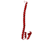
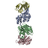
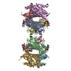
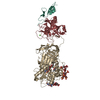
| Title | Crystal Structure of Reovirus Attachment Protein Sigma1 Trimer | ||||||
 Components Components | SIGMA 1 PROTEIN | ||||||
 Keywords Keywords | VIRAL PROTEIN / reovirus / sigma1 / fiber / beta-spiral / beta-barrel / trimer / receptor-binding | ||||||
| Function / homology |  Function and homology information Function and homology informationviral outer capsid / cell adhesion / symbiont entry into host cell / virion attachment to host cell Similarity search - Function | ||||||
| Biological species |  Mammalian orthoreovirus 3 Mammalian orthoreovirus 3 | ||||||
| Method |  X-RAY DIFFRACTION / MIR / Resolution: 2.6 Å X-RAY DIFFRACTION / MIR / Resolution: 2.6 Å | ||||||
 Authors Authors | Chappell, J.D. / Prota, A.E. / Dermody, T.S. / Stehle, T. | ||||||
 Citation Citation | Journal: EMBO J. / Year: 2002 Title: Crystal structure of reovirus attachment protein sigma1 reveals evolutionary relationship to adenovirus fiber. Authors: Chappell, J.D. / Prota, A.E. / Dermody, T.S. / Stehle, T. | ||||||
| History |
|
- Structure visualization
| Structure viewer | Molecule: MolmilJmol/JSmol |
|---|
- Downloads & links
-Download
| PDBx/mmCIF format | 1kke.cif.gz | 127.9 KB | Display | PDBx/mmCIF format |
|---|---|---|---|---|
| PDB format | pdb1kke.ent.gz | 101 KB | Display | PDB format |
| PDBx/mmJSON format | 1kke.json.gz | Tree view | PDBx/mmJSON format | |
| Others |  Other downloads Other downloads |
-Validation report
| Summary document | 1kke_validation.pdf.gz | 438.3 KB | Display | wwPDB validaton report |
|---|---|---|---|---|
| Full document | 1kke_full_validation.pdf.gz | 445.9 KB | Display | |
| Data in XML | 1kke_validation.xml.gz | 25 KB | Display | |
| Data in CIF | 1kke_validation.cif.gz | 34 KB | Display | |
| Arichive directory | https://data.pdbj.org/pub/pdb/validation_reports/kk/1kkeftp://data.pdbj.org/pub/pdb/validation_reports/kk/1kke | HTTPS FTP |
-Related structure data
| Similar structure data |
|---|


-Links
 PDBj
PDBj
- Assembly
| Deposited unit | 
| ||||||||
|---|---|---|---|---|---|---|---|---|---|
| 1 |
| ||||||||
| Unit cell |
| ||||||||
| Details | The asymmetric unit contains a biologically active trimer |
-Components
| #1: Protein | Mass: 22687.412 Da / Num. of mol.: 3 Source method: isolated from a genetically manipulated source Source: (gene. exp.) Mammalian orthoreovirus 3 / Genus: Orthoreovirus / Species: Mammalian orthoreovirus / Strain: type 3 strain dearing / Cell line (production host): SF21 / Production host:   Spodoptera frugiperda (fall armyworm) / References: UniProt: P03528 Spodoptera frugiperda (fall armyworm) / References: UniProt: P03528#2: Water | ChemComp-HOH / |  Mass: 18.015 Da / Num. of mol.: 189 / Source method: isolated from a natural source / Formula: H2O Mass: 18.015 Da / Num. of mol.: 189 / Source method: isolated from a natural source / Formula: H2O |
|---|
-Experimental details
-Experiment
| Experiment | Method: X-RAY DIFFRACTION / Number of used crystals: 2 |
|---|
- Sample preparation
| Crystal | Density Matthews: 2.06 Å3/Da / Density % sol: 40.37 % | ||||||||||||||||||||||||||||||
|---|---|---|---|---|---|---|---|---|---|---|---|---|---|---|---|---|---|---|---|---|---|---|---|---|---|---|---|---|---|---|---|
| Crystal grow | Temperature: 293 K / Method: vapor diffusion, hanging drop / pH: 7.5 Details: PEG4000, Isopropanol, Hepes, pH 7.5, VAPOR DIFFUSION, HANGING DROP, temperature 293K | ||||||||||||||||||||||||||||||
| Crystal grow | *PLUS Temperature: 20 ℃ / Method: vapor diffusion | ||||||||||||||||||||||||||||||
| Components of the solutions | *PLUS
|
-Data collection
| Diffraction | Mean temperature: 293 K |
|---|---|
| Diffraction source | Source: ROTATING ANODE / Type: RIGAKU RU300 / Wavelength: 1.541789 Å |
| Detector | Type: RIGAKU RAXIS IV / Detector: IMAGE PLATE / Date: Feb 1, 2001 / Details: mirror |
| Radiation | Protocol: SINGLE WAVELENGTH / Monochromatic (M) / Laue (L): M / Scattering type: x-ray |
| Radiation wavelength | Wavelength: 1.541789 Å / Relative weight: 1 |
| Reflection | Resolution: 2.6→20 Å / Num. all: 17954 / Num. obs: 16338 / % possible obs: 91 % / Observed criterion σ(F): 2 / Observed criterion σ(I): 2 / Rmerge(I) obs: 0.087 / Net I/σ(I): 9 |
| Reflection shell | Resolution: 2.6→2.7 Å / Rmerge(I) obs: 0.284 / Mean I/σ(I) obs: 2.2 / % possible all: 64 |
| Reflection | *PLUS Lowest resolution: 20 Å / % possible obs: 91 % / Num. measured all: 56260 |
| Reflection shell | *PLUS % possible obs: 64 % |
- Processing
| Software |
| ||||||||||||||||||||
|---|---|---|---|---|---|---|---|---|---|---|---|---|---|---|---|---|---|---|---|---|---|
| Refinement | Method to determine structure: MIR / Resolution: 2.6→20 Å / σ(F): 1 Stereochemistry target values: xplor parameter set (Engh & Huber)
| ||||||||||||||||||||
| Refinement step | Cycle: LAST / Resolution: 2.6→20 Å
| ||||||||||||||||||||
| Refine LS restraints |
| ||||||||||||||||||||
| Software | *PLUS Name: X-PLOR / Version: 3.1 / Classification: refinement | ||||||||||||||||||||
| Refinement | *PLUS Highest resolution: 2.6 Å / Lowest resolution: 20 Å / σ(F): 1 / % reflection Rfree: 5 % / Rfactor all: 0.172 / Rfactor Rfree: 0.235 | ||||||||||||||||||||
| Solvent computation | *PLUS | ||||||||||||||||||||
| Displacement parameters | *PLUS | ||||||||||||||||||||
| Refine LS restraints | *PLUS Type: x_angle_deg / Dev ideal: 1.4 |
