 Movie
Movie Controller


+ Open data

- Basic information
| Entry | Database: PDB / ID: 1kkc | ||||||
|---|---|---|---|---|---|---|---|
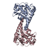
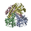
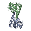
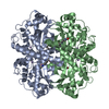
| Title | Crystal structure of Aspergillus fumigatus MnSOD | ||||||
 Components Components | Manganese Superoxide Dismutase | ||||||
 Keywords Keywords | OXIDOREDUCTASE / Homotetramer | ||||||
| Function / homology |  Function and homology information Function and homology informationIgE binding / superoxide dismutase / superoxide dismutase activity / manganese ion binding / mitochondrial matrix / mitochondrion Similarity search - Function | ||||||
| Biological species |  | ||||||
| Method |  X-RAY DIFFRACTION / MOLECULAR REPLACEMENT / Resolution: 2 Å X-RAY DIFFRACTION / MOLECULAR REPLACEMENT / Resolution: 2 Å | ||||||
 Authors Authors | Fluckiger, S. / Mittl, P.R.E. / Scapozza, L. / Fijten, H. / Folkers, G. / Grutter, M.G. / Blaser, K. / Crameri, R. | ||||||
 Citation Citation | Journal: J.Immunol. / Year: 2002 Title: Comparison of the crystal structures of the human manganese superoxide dismutase and the homologous Aspergillus fumigatus allergen at 2-A resolution. Authors: Fluckiger, S. / Mittl, P.R. / Scapozza, L. / Fijten, H. / Folkers, G. / Grutter, M.G. / Blaser, K. / Crameri, R. | ||||||
| History |
|
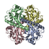
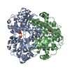
- Structure visualization
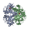
| Structure viewer | Molecule: MolmilJmol/JSmol |
|---|
- Downloads & links
-Download
| PDBx/mmCIF format | 1kkc.cif.gz | 181.5 KB | Display | PDBx/mmCIF format |
|---|---|---|---|---|
| PDB format | pdb1kkc.ent.gz | 143.9 KB | Display | PDB format |
| PDBx/mmJSON format | 1kkc.json.gz | Tree view | PDBx/mmJSON format | |
| Others |  Other downloads Other downloads |
-Validation report
| Arichive directory | https://data.pdbj.org/pub/pdb/validation_reports/kk/1kkcftp://data.pdbj.org/pub/pdb/validation_reports/kk/1kkc | HTTPS FTP |
|---|
-Related structure data
| Similar structure data |
|---|

-Links
 PDBj
PDBj
- Assembly
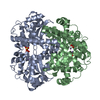
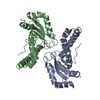
| Deposited unit | 
| ||||||||
|---|---|---|---|---|---|---|---|---|---|
| 1 |
| ||||||||
| Unit cell |
|
-Components
| #1: Protein | Mass: 24516.637 Da / Num. of mol.: 4 Source method: isolated from a genetically manipulated source Source: (gene. exp.)  #2: Chemical | ChemComp-MN /   Mass: 54.938 Da / Num. of mol.: 4 / Source method: obtained synthetically / Formula: Mn Mass: 54.938 Da / Num. of mol.: 4 / Source method: obtained synthetically / Formula: Mn#3: Water | ChemComp-HOH / |  Mass: 18.015 Da / Num. of mol.: 745 / Source method: isolated from a natural source / Formula: H2O Mass: 18.015 Da / Num. of mol.: 745 / Source method: isolated from a natural source / Formula: H2O |
|---|
-Experimental details
-Experiment
| Experiment | Method: X-RAY DIFFRACTION / Number of used crystals: 1 |
|---|
- Sample preparation
| Crystal | Density Matthews: 2.31 Å3/Da / Density % sol: 46.69 % | |||||||||||||||||||||||||||||||||||
|---|---|---|---|---|---|---|---|---|---|---|---|---|---|---|---|---|---|---|---|---|---|---|---|---|---|---|---|---|---|---|---|---|---|---|---|---|
| Crystal grow | Temperature: 296 K / Method: vapor diffusion, hanging drop / pH: 8 Details: PEG 400, pH 8.0, VAPOR DIFFUSION, HANGING DROP, temperature 296K | |||||||||||||||||||||||||||||||||||
| Crystal grow | *PLUS Temperature: 23 ℃ / pH: 8 | |||||||||||||||||||||||||||||||||||
| Components of the solutions | *PLUS
|
-Data collection
| Diffraction | Mean temperature: 100 K |
|---|---|
| Diffraction source | Source: ROTATING ANODE / Type: ENRAF-NONIUS FR591 / Wavelength: 1.5418 |
| Detector | Type: Prophysics XRM-216 / Detector: IMAGE PLATE / Date: Jun 23, 2000 / Details: double-focusing mirror system |
| Radiation | Monochromator: ProPhysics MIRRORS / Protocol: SINGLE WAVELENGTH / Monochromatic (M) / Laue (L): M / Scattering type: x-ray |
| Radiation wavelength | Wavelength: 1.5418 Å / Relative weight: 1 |
| Reflection | Resolution: 2→50 Å / Num. obs: 56622 / % possible obs: 91 % / Observed criterion σ(F): 1 / Observed criterion σ(I): 1 |
| Reflection shell | Resolution: 2→2.07 Å / % possible all: 34.8 |
| Reflection | *PLUS Lowest resolution: 50 Å / Num. obs: 56585 / Num. measured all: 257369 / Rmerge(I) obs: 0.083 |
| Reflection shell | *PLUS % possible obs: 34.8 % / Rmerge(I) obs: 0.333 |
- Processing
| Software |
| ||||||||||||||||
|---|---|---|---|---|---|---|---|---|---|---|---|---|---|---|---|---|---|
| Refinement | Method to determine structure: MOLECULAR REPLACEMENT Starting model: homology model Resolution: 2→50 Å / σ(F): 0 / Stereochemistry target values: Engh & Huber
| ||||||||||||||||
| Displacement parameters |
| ||||||||||||||||
| Refinement step | Cycle: LAST / Resolution: 2→50 Å
| ||||||||||||||||
| Refine LS restraints |
| ||||||||||||||||
| Software | *PLUS Name: CNS / Classification: refinement | ||||||||||||||||
| Refinement | *PLUS Highest resolution: 2 Å / Lowest resolution: 500 Å / σ(F): 0 / % reflection Rfree: 10 % / Rfactor obs: 0.194 | ||||||||||||||||
| Solvent computation | *PLUS | ||||||||||||||||
| Displacement parameters | *PLUS |
