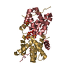
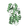
 Movie
Movie Controller


+ Open data

- Basic information
| Entry | Database: PDB / ID: 1kjn | ||||||
|---|---|---|---|---|---|---|---|
| Title | Structure of MT0777 | ||||||
 Components Components | MTH0777 | ||||||
 Keywords Keywords | STRUCTURAL GENOMICS / UNKNOWN FUNCTION / hypotethical protein / PSI / Protein Structure Initiative / Midwest Center for Structural Genomics / MCSG | ||||||
| Function / homology | MTH777-like / Uncharacterised conserved protein UCP006600 / MTH777-like superfamily / Domain of unknown function (DUF1890) / Rossmann fold / 3-Layer(aba) Sandwich / Alpha Beta / Conserved protein Function and homology information Function and homology information | ||||||
| Biological species |   Methanothermobacter thermautotrophicus (archaea) Methanothermobacter thermautotrophicus (archaea) | ||||||
| Method |  X-RAY DIFFRACTION / SYNCHROTRON / MAD / Resolution: 2.2 Å X-RAY DIFFRACTION / SYNCHROTRON / MAD / Resolution: 2.2 Å | ||||||
 Authors Authors | Christendat, D. / Edwards, A. / Joachimiak, A. / Korolev, S. / Midwest Center for Structural Genomics (MCSG) | ||||||
 Citation Citation | Journal: To be published Title: Crystal structure of MT0777 Authors: Christendat, D. / Edwards, A. / Joachimiak, A. / Korolev, S. | ||||||
| History |
|
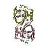
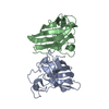
- Structure visualization
| Structure viewer | Molecule: MolmilJmol/JSmol |
|---|
- Downloads & links
-Download
| PDBx/mmCIF format | 1kjn.cif.gz | 72.8 KB | Display | PDBx/mmCIF format |
|---|---|---|---|---|
| PDB format | pdb1kjn.ent.gz | 55.4 KB | Display | PDB format |
| PDBx/mmJSON format | 1kjn.json.gz | Tree view | PDBx/mmJSON format | |
| Others |  Other downloads Other downloads |
-Validation report
| Summary document | 1kjn_validation.pdf.gz | 435.3 KB | Display | wwPDB validaton report |
|---|---|---|---|---|
| Full document | 1kjn_full_validation.pdf.gz | 440.8 KB | Display | |
| Data in XML | 1kjn_validation.xml.gz | 17.6 KB | Display | |
| Data in CIF | 1kjn_validation.cif.gz | 23.1 KB | Display | |
| Arichive directory | https://data.pdbj.org/pub/pdb/validation_reports/kj/1kjnftp://data.pdbj.org/pub/pdb/validation_reports/kj/1kjn | HTTPS FTP |
-Related structure data
| Similar structure data | |
|---|---|
| Other databases |


-Links
 PDBj
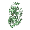
PDBj- Assembly
| Deposited unit | 
| ||||||||
|---|---|---|---|---|---|---|---|---|---|
| 1 |
| ||||||||
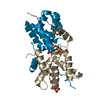
| Unit cell |
|
-Components
| #1: Protein | Mass: 17479.367 Da / Num. of mol.: 2 / Fragment: hypothetical protein Source method: isolated from a genetically manipulated source Source: (gene. exp.) Methanothermobacter thermautotrophicus (archaea)Production host:  #2: Water | ChemComp-HOH / |  Mass: 18.015 Da / Num. of mol.: 173 / Source method: isolated from a natural source / Formula: H2O Mass: 18.015 Da / Num. of mol.: 173 / Source method: isolated from a natural source / Formula: H2OHas protein modification | Y | |
|---|
-Experimental details
-Experiment
| Experiment | Method: X-RAY DIFFRACTION / Number of used crystals: 1 |
|---|
- Sample preparation
| Crystal | Density Matthews: 2.32 Å3/Da / Density % sol: 46.93 % |
|---|---|
| Crystal grow | Temperature: 298 K / Method: vapor diffusion, hanging drop / pH: 7.5 Details: PEG 3350, pH 7.5, VAPOR DIFFUSION, HANGING DROP, temperature 298.0K |
-Data collection
| Diffraction | Mean temperature: 100 K | ||||||||||||
|---|---|---|---|---|---|---|---|---|---|---|---|---|---|
| Diffraction source | Source: SYNCHROTRON / Site: APS  / Beamline: 19-ID / Wavelength: 0.97938, 0.97916, 0.97779 / Beamline: 19-ID / Wavelength: 0.97938, 0.97916, 0.97779 | ||||||||||||
| Detector | Type: SBC-1 / Detector: CCD / Date: Jul 8, 2001 | ||||||||||||
| Radiation | Monochromator: SAGITALLY FOCUSED Si(111) / Protocol: MAD / Monochromatic (M) / Laue (L): M / Scattering type: x-ray | ||||||||||||
| Radiation wavelength |
| ||||||||||||
| Reflection | Resolution: 2→50 Å / Num. all: 42424 / Num. obs: 37502 / % possible obs: 88.4 % / Observed criterion σ(I): -3 / Redundancy: 4 % / Biso Wilson estimate: 20.2 Å2 / Rmerge(I) obs: 0.066 / Net I/σ(I): 16 | ||||||||||||
| Reflection shell | Resolution: 2→2.07 Å / Redundancy: 1 % / Rmerge(I) obs: 0.553 / Mean I/σ(I) obs: 1.4 / % possible all: 40.3 |
- Processing
| Software |
| ||||||||||||||||||||||||||||||||||||||||||||||||||||||||||||||||||||||||||||||||
|---|---|---|---|---|---|---|---|---|---|---|---|---|---|---|---|---|---|---|---|---|---|---|---|---|---|---|---|---|---|---|---|---|---|---|---|---|---|---|---|---|---|---|---|---|---|---|---|---|---|---|---|---|---|---|---|---|---|---|---|---|---|---|---|---|---|---|---|---|---|---|---|---|---|---|---|---|---|---|---|---|---|
| Refinement | Method to determine structure: MAD / Resolution: 2.2→48.07 Å / Rfactor Rfree error: 0.009 / Data cutoff high absF: 1858443.3 / Data cutoff high rms absF: 1858443.3 / Data cutoff low absF: 0 / Isotropic thermal model: RESTRAINED / Cross valid method: THROUGHOUT / σ(F): 0 / Stereochemistry target values: Engh & Huber
| ||||||||||||||||||||||||||||||||||||||||||||||||||||||||||||||||||||||||||||||||
| Solvent computation | Solvent model: FLAT MODEL / Bsol: 24.0748 Å2 / ksol: 0.344549 e/Å3 | ||||||||||||||||||||||||||||||||||||||||||||||||||||||||||||||||||||||||||||||||
| Displacement parameters | Biso mean: 31.5 Å2
| ||||||||||||||||||||||||||||||||||||||||||||||||||||||||||||||||||||||||||||||||
| Refine analyze |
| ||||||||||||||||||||||||||||||||||||||||||||||||||||||||||||||||||||||||||||||||
| Refinement step | Cycle: LAST / Resolution: 2.2→48.07 Å
| ||||||||||||||||||||||||||||||||||||||||||||||||||||||||||||||||||||||||||||||||
| Refine LS restraints |
| ||||||||||||||||||||||||||||||||||||||||||||||||||||||||||||||||||||||||||||||||
| LS refinement shell | Resolution: 2.2→2.28 Å / Rfactor Rfree error: 0.026 / Total num. of bins used: 6
| ||||||||||||||||||||||||||||||||||||||||||||||||||||||||||||||||||||||||||||||||
| Xplor file |
|
