 Movie
Movie Controller

[English] 日本語
 Yorodumi
- PDB-1kj8: Crystal Structure of PurT-Encoded Glycinamide Ribonucleotide Tran... -
+ Open data

- Basic information
| Entry | Database: PDB / ID: 1kj8 | ||||||
|---|---|---|---|---|---|---|---|
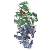
| Title | Crystal Structure of PurT-Encoded Glycinamide Ribonucleotide Transformylase in Complex with Mg-ATP and GAR | ||||||
 Components Components | phosphoribosylglycinamide formyltransferase 2 | ||||||
 Keywords Keywords | TRANSFERASE / ATP-grasp / purine biosynthesis / nucleotide | ||||||
| Function / homology |  Function and homology information Function and homology informationphosphoribosylglycinamide formyltransferase 2 / phosphoribosylglycinamide formyltransferase 2 activity / acetate kinase activity / phosphoribosylglycinamide formyltransferase activity / 'de novo' IMP biosynthetic process / magnesium ion binding / ATP binding / cytosol Similarity search - Function | ||||||
| Biological species |  | ||||||
| Method |  X-RAY DIFFRACTION / FOURIER SYNTHESIS / Resolution: 1.6 Å X-RAY DIFFRACTION / FOURIER SYNTHESIS / Resolution: 1.6 Å | ||||||
 Authors Authors | Thoden, J.B. / Firestine, S.M. / Benkovic, S.J. / Holden, H.M. | ||||||
 Citation Citation | Journal: J.Biol.Chem. / Year: 2002 Title: PurT-encoded glycinamide ribonucleotide transformylase. Accommodation of adenosine nucleotide analogs within the active site. Authors: Thoden, J.B. / Firestine, S.M. / Benkovic, S.J. / Holden, H.M. | ||||||
| History |
|
- Structure visualization
| Structure viewer | Molecule: MolmilJmol/JSmol |
|---|
- Downloads & links
-Download
| PDBx/mmCIF format | 1kj8.cif.gz | 186.2 KB | Display | PDBx/mmCIF format |
|---|---|---|---|---|
| PDB format | pdb1kj8.ent.gz | 143.4 KB | Display | PDB format |
| PDBx/mmJSON format | 1kj8.json.gz | Tree view | PDBx/mmJSON format | |
| Others |  Other downloads Other downloads |
-Validation report
| Summary document | 1kj8_validation.pdf.gz | 1.7 MB | Display | wwPDB validaton report |
|---|---|---|---|---|
| Full document | 1kj8_full_validation.pdf.gz | 1.7 MB | Display | |
| Data in XML | 1kj8_validation.xml.gz | 42 KB | Display | |
| Data in CIF | 1kj8_validation.cif.gz | 63.5 KB | Display | |
| Arichive directory | https://data.pdbj.org/pub/pdb/validation_reports/kj/1kj8ftp://data.pdbj.org/pub/pdb/validation_reports/kj/1kj8 | HTTPS FTP |
-Related structure data
| Related structure data | 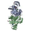 1kj9C 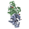 1kjiC 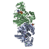 1kjjC 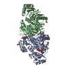 1kjqC 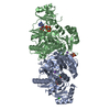 1eyzS S: Starting model for refinement C: citing same article ( |
|---|---|
| Similar structure data |


-Links
 PDBj
PDBj


- Assembly
| Deposited unit | 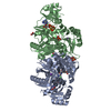
| ||||||||||
|---|---|---|---|---|---|---|---|---|---|---|---|
| 1 |
| ||||||||||
| Unit cell |
|
-Components
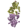
-Protein , 1 types, 2 molecules AB
| #1: Protein | Mass: 42349.340 Da / Num. of mol.: 2 Source method: isolated from a genetically manipulated source Source: (gene. exp.) References: UniProt: P33221, Transferases; Transferring one-carbon groups; Hydroxymethyl-, formyl- and related transferases |
|---|
-Non-polymers , 8 types, 864 molecules 


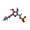


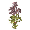
| #2: Chemical | ChemComp-MG /  Mass: 24.305 Da / Num. of mol.: 4 / Source method: obtained synthetically / Formula: Mg Mass: 24.305 Da / Num. of mol.: 4 / Source method: obtained synthetically / Formula: Mg#3: Chemical |  Mass: 22.990 Da / Num. of mol.: 2 / Source method: obtained synthetically / Formula: Na Mass: 22.990 Da / Num. of mol.: 2 / Source method: obtained synthetically / Formula: Na#4: Chemical | ChemComp-CL /  Mass: 35.453 Da / Num. of mol.: 4 / Source method: obtained synthetically / Formula: Cl Mass: 35.453 Da / Num. of mol.: 4 / Source method: obtained synthetically / Formula: Cl#5: Chemical |  Mass: 507.181 Da / Num. of mol.: 2 / Source method: obtained synthetically / Formula: C10H16N5O13P3 / Comment: ATP, energy-carrying molecule*YM Mass: 507.181 Da / Num. of mol.: 2 / Source method: obtained synthetically / Formula: C10H16N5O13P3 / Comment: ATP, energy-carrying molecule*YM#6: Chemical |  Mass: 284.160 Da / Num. of mol.: 2 / Source method: obtained synthetically / Formula: C7H13N2O8P Mass: 284.160 Da / Num. of mol.: 2 / Source method: obtained synthetically / Formula: C7H13N2O8P#7: Chemical | ChemComp-MPO / |  Mass: 209.263 Da / Num. of mol.: 1 / Source method: obtained synthetically / Formula: C7H15NO4S / Comment: pH buffer*YM Mass: 209.263 Da / Num. of mol.: 1 / Source method: obtained synthetically / Formula: C7H15NO4S / Comment: pH buffer*YM#8: Chemical |  Mass: 62.068 Da / Num. of mol.: 3 / Source method: obtained synthetically / Formula: C2H6O2 Mass: 62.068 Da / Num. of mol.: 3 / Source method: obtained synthetically / Formula: C2H6O2#9: Water | ChemComp-HOH / | Mass: 18.015 Da / Num. of mol.: 846 / Source method: isolated from a natural source / Formula: H2O |
|---|
-Experimental details
-Experiment
| Experiment | Method: X-RAY DIFFRACTION / Number of used crystals: 1 |
|---|
- Sample preparation
| Crystal | Density Matthews: 2.49 Å3/Da / Density % sol: 50.61 % | ||||||||||||||||||||||||||||||||||||||||||||||||||||||||
|---|---|---|---|---|---|---|---|---|---|---|---|---|---|---|---|---|---|---|---|---|---|---|---|---|---|---|---|---|---|---|---|---|---|---|---|---|---|---|---|---|---|---|---|---|---|---|---|---|---|---|---|---|---|---|---|---|---|
| Crystal grow | Temperature: 277 K / Method: batch / pH: 6.7 Details: PEG 5000, NaCl, MgCl2, MOPS, ATP, GAR, pH 6.7, batch at 277K | ||||||||||||||||||||||||||||||||||||||||||||||||||||||||
| Crystal grow | *PLUS Temperature: 4 ℃ / Method: batch method / Details: used macroseeding | ||||||||||||||||||||||||||||||||||||||||||||||||||||||||
| Components of the solutions | *PLUS
|
-Data collection
| Diffraction | Mean temperature: 110 K |
|---|---|
| Diffraction source | Source: ROTATING ANODE / Type: RIGAKU RU200 / Wavelength: 1.5418 Å |
| Detector | Type: SIEMENS HI-STAR / Detector: AREA DETECTOR / Date: Jun 7, 2000 / Details: goebel mirrors |
| Radiation | Monochromator: goebel optics / Protocol: SINGLE WAVELENGTH / Monochromatic (M) / Laue (L): M / Scattering type: x-ray |
| Radiation wavelength | Wavelength: 1.5418 Å / Relative weight: 1 |
| Reflection | Resolution: 1.6→30 Å / Num. all: 108032 / Num. obs: 108032 / % possible obs: 97 % / Observed criterion σ(F): 0 / Observed criterion σ(I): 0 / Redundancy: 3.1 % / Rmerge(I) obs: 0.044 / Net I/σ(I): 18.9 |
| Reflection shell | Resolution: 1.6→1.67 Å / Redundancy: 2 % / Rmerge(I) obs: 0.212 / Mean I/σ(I) obs: 3.4 / Num. unique all: 12764 / % possible all: 92 |
| Reflection | *PLUS Rmerge(I) obs: 0.044 |
| Reflection shell | *PLUS % possible obs: 92 % / Num. unique obs: 12764 / Rmerge(I) obs: 0.212 |
- Processing
| Software |
| |||||||||||||||||||||||||
|---|---|---|---|---|---|---|---|---|---|---|---|---|---|---|---|---|---|---|---|---|---|---|---|---|---|---|
| Refinement | Method to determine structure: FOURIER SYNTHESIS Starting model: PDB ENTRY 1EYZ Resolution: 1.6→30 Å / Isotropic thermal model: Isotropic / Cross valid method: THROUGHOUT / σ(F): 0 / Stereochemistry target values: Engh & Huber
| |||||||||||||||||||||||||
| Refinement step | Cycle: LAST / Resolution: 1.6→30 Å
| |||||||||||||||||||||||||
| Refine LS restraints |
| |||||||||||||||||||||||||
| Refinement | *PLUS Rfactor all: 0.182 / Rfactor Rfree: 0.228 / Rfactor Rwork: 0.181 | |||||||||||||||||||||||||
| Solvent computation | *PLUS | |||||||||||||||||||||||||
| Displacement parameters | *PLUS | |||||||||||||||||||||||||
| Refine LS restraints | *PLUS
|
