 Movie
Movie Controller

[English] 日本語
 Yorodumi
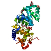
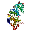
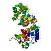
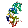
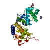
- PDB-1kg5: Crystal structure of the K142Q mutant of E.coli MutY (core fragment) -
+ Open data

- Basic information
| Entry | Database: PDB / ID: 1kg5 | ||||||
|---|---|---|---|---|---|---|---|
| Title | Crystal structure of the K142Q mutant of E.coli MutY (core fragment) | ||||||
 Components Components | A/G-specific adenine glycosylase | ||||||
 Keywords Keywords | HYDROLASE / DNA Repair | ||||||
| Function / homology |  Function and homology information Function and homology informationadenine/guanine mispair binding / adenine glycosylase / 8-oxo-7,8-dihydroguanine DNA N-glycosylase activity / purine-specific mismatch base pair DNA N-glycosylase activity / oxidized purine DNA binding / mismatch repair / base-excision repair / 4 iron, 4 sulfur cluster binding / metal ion binding Similarity search - Function | ||||||
| Biological species |  | ||||||
| Method |  X-RAY DIFFRACTION / SYNCHROTRON / Difference fourier / Resolution: 1.35 Å X-RAY DIFFRACTION / SYNCHROTRON / Difference fourier / Resolution: 1.35 Å | ||||||
 Authors Authors | Gilboa, R. / Kilshtein, A. / Zharkov, D.O. / Kycia, J.H. / Gerchman, S.E. / Grollman, A.P. / Shoham, G. | ||||||
 Citation Citation | Journal: To be Published Title: Analysis of the E.coli MutY DNA glycosylase structure and function by site-directed mutagenesis Authors: Gilboa, R. / Kilshtein, A. / Zharkov, D.O. / Kycia, J.H. / Gerchman, S.E. / Grollman, A.P. / Shoham, G. #1: Journal: Biochemistry / Year: 2000Title: Role for Lysine 142 in the excision of adenine from A:G mispairs by MutY DNA glycosylase of Escherichia coli. Authors: Zharkov, D.O. / Gilboa, R. / Yagil, I. / Kycia, J.H. / Gerchman, S.E. / Shoham, G. / Grollmam, A.P. | ||||||
| History |
|
- Structure visualization
| Structure viewer | Molecule: MolmilJmol/JSmol |
|---|
- Downloads & links
-Download
| PDBx/mmCIF format | 1kg5.cif.gz | 117.8 KB | Display | PDBx/mmCIF format |
|---|---|---|---|---|
| PDB format | pdb1kg5.ent.gz | 90 KB | Display | PDB format |
| PDBx/mmJSON format | 1kg5.json.gz | Tree view | PDBx/mmJSON format | |
| Others |  Other downloads Other downloads |
-Validation report
| Summary document | 1kg5_validation.pdf.gz | 399.1 KB | Display | wwPDB validaton report |
|---|---|---|---|---|
| Full document | 1kg5_full_validation.pdf.gz | 401.4 KB | Display | |
| Data in XML | 1kg5_validation.xml.gz | 6.4 KB | Display | |
| Data in CIF | 1kg5_validation.cif.gz | 11.1 KB | Display | |
| Arichive directory | https://data.pdbj.org/pub/pdb/validation_reports/kg/1kg5ftp://data.pdbj.org/pub/pdb/validation_reports/kg/1kg5 | HTTPS FTP |
-Related structure data
| Related structure data |  1kg2SC  1kg3C  1kg4C  1kg6C  1kg7C S: Starting model for refinement C: citing same article ( |
|---|---|
| Similar structure data |
-Links
 PDBj
PDBj

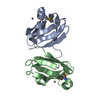
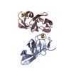
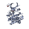
- Assembly
| Deposited unit | 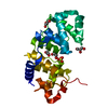
| |||||||||
|---|---|---|---|---|---|---|---|---|---|---|
| 1 |
| |||||||||
| Unit cell |
| |||||||||
| Components on special symmetry positions |
|
-Components
| #1: Protein | Mass: 25047.939 Da / Num. of mol.: 1 / Fragment: Catalytic domain / Mutation: K142Q Source method: isolated from a genetically manipulated source Source: (gene. exp.) References: UniProt: P17802, Hydrolases; Glycosylases; Hydrolysing N-glycosyl compounds | ||
|---|---|---|---|
| #2: Chemical | ChemComp-SO4 /   Mass: 96.063 Da / Num. of mol.: 1 / Source method: obtained synthetically / Formula: SO4 Mass: 96.063 Da / Num. of mol.: 1 / Source method: obtained synthetically / Formula: SO4 | ||
| #3: Chemical | ChemComp-SF4 /   Mass: 351.640 Da / Num. of mol.: 1 / Source method: obtained synthetically / Formula: Fe4S4 Mass: 351.640 Da / Num. of mol.: 1 / Source method: obtained synthetically / Formula: Fe4S4 | ||
| #4: Chemical | ChemComp-GOL /   Mass: 92.094 Da / Num. of mol.: 4 / Source method: obtained synthetically / Formula: C3H8O3 Mass: 92.094 Da / Num. of mol.: 4 / Source method: obtained synthetically / Formula: C3H8O3#5: Water | ChemComp-HOH / |  Mass: 18.015 Da / Num. of mol.: 309 / Source method: isolated from a natural source / Formula: H2O Mass: 18.015 Da / Num. of mol.: 309 / Source method: isolated from a natural source / Formula: H2O |
-Experimental details
-Experiment
| Experiment | Method: X-RAY DIFFRACTION / Number of used crystals: 1 |
|---|
- Sample preparation
| Crystal | Density Matthews: 1.75 Å3/Da / Density % sol: 50.26 % |
|---|---|
| Crystal grow | Temperature: 288 K / Method: vapor diffusion, hanging drop / pH: 8 Details: Lithium sulfate, HEPES, Sodium chloride, Magnesium sulfate, pH 8.0, VAPOR DIFFUSION, HANGING DROP, temperature 288K |
-Data collection
| Diffraction | Mean temperature: 100 K |
|---|---|
| Diffraction source | Source: SYNCHROTRON / Site: NSLS  / Beamline: X26C / Wavelength: 1.1 Å / Beamline: X26C / Wavelength: 1.1 Å |
| Detector | Type: ADSC QUANTUM 4 / Detector: CCD / Date: Sep 16, 1999 |
| Radiation | Protocol: SINGLE WAVELENGTH / Monochromatic (M) / Laue (L): M / Scattering type: x-ray |
| Radiation wavelength | Wavelength: 1.1 Å / Relative weight: 1 |
| Reflection | Resolution: 1.35→40 Å / Num. all: 49330 / Num. obs: 49330 / % possible obs: 91.7 % / Observed criterion σ(I): 0 / Redundancy: 4.5 % / Biso Wilson estimate: 11.1 Å2 / Rsym value: 0.062 / Net I/σ(I): 13.5 |
| Reflection shell | Resolution: 1.35→1.37 Å / Redundancy: 2 % / Num. unique all: 1464 / Rsym value: 0.214 / % possible all: 55.4 |
- Processing
| Software |
| |||||||||||||||||||||||||||||||||
|---|---|---|---|---|---|---|---|---|---|---|---|---|---|---|---|---|---|---|---|---|---|---|---|---|---|---|---|---|---|---|---|---|---|---|
| Refinement | Method to determine structure: Difference fourier Starting model: 1KG2 Resolution: 1.35→40 Å / Num. parameters: 19273 / Num. restraintsaints: 23153 / Cross valid method: FREE R / σ(F): 0 / Stereochemistry target values: ENGH & HUBER / Details: CNS 0.9 WAS ALSO USED IN REFINEMENT.
| |||||||||||||||||||||||||||||||||
| Displacement parameters | Biso mean: 15.5 Å2 | |||||||||||||||||||||||||||||||||
| Refine analyze | Luzzati coordinate error obs: 0.11 Å / Num. disordered residues: 9 / Occupancy sum hydrogen: 0 / Occupancy sum non hydrogen: 2110 | |||||||||||||||||||||||||||||||||
| Refinement step | Cycle: LAST / Resolution: 1.35→40 Å
| |||||||||||||||||||||||||||||||||
| Refine LS restraints |
|
