 Movie
Movie Controller

[English] 日本語
 Yorodumi
- PDB-1kfm: Core side-chain packing and backbone conformation in Lpp-56 coile... -
+ Open data

- Basic information
| Entry | Database: PDB / ID: 1kfm | ||||||
|---|---|---|---|---|---|---|---|
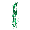
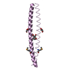
| Title | Core side-chain packing and backbone conformation in Lpp-56 coiled-coil mutants | ||||||
 Components Components | MAJOR OUTER MEMBRANE LIPOPROTEIN | ||||||
 Keywords Keywords | MEMBRANE PROTEIN / LIPOPROTEIN / PROTEIN FOLDING / HELIX CAPPING / ALANINE-ZIPPER | ||||||
| Function / homology |  Function and homology information Function and homology informationperiplasmic space organization / lipid modification / peptidoglycan binding / cell outer membrane / outer membrane-bounded periplasmic space / lipid binding / membrane / identical protein binding Similarity search - Function | ||||||
| Biological species |  | ||||||
| Method |  X-RAY DIFFRACTION / SYNCHROTRON / MOLECULAR REPLACEMENT / Resolution: 2 Å X-RAY DIFFRACTION / SYNCHROTRON / MOLECULAR REPLACEMENT / Resolution: 2 Å | ||||||
 Authors Authors | Liu, J. / Cao, W. / Lu, M. | ||||||
 Citation Citation | Journal: J.Mol.Biol. / Year: 2002 Title: Core side-chain packing and backbone conformation in Lpp-56 coiled-coil mutants. Authors: Liu, J. / Cao, W. / Lu, M. | ||||||
| History |
|
- Structure visualization
| Structure viewer | Molecule: MolmilJmol/JSmol |
|---|
- Downloads & links
-Download
| PDBx/mmCIF format | 1kfm.cif.gz | 19.8 KB | Display | PDBx/mmCIF format |
|---|---|---|---|---|
| PDB format | pdb1kfm.ent.gz | 12.2 KB | Display | PDB format |
| PDBx/mmJSON format | 1kfm.json.gz | Tree view | PDBx/mmJSON format | |
| Others |  Other downloads Other downloads |
-Validation report
| Arichive directory | https://data.pdbj.org/pub/pdb/validation_reports/kf/1kfmftp://data.pdbj.org/pub/pdb/validation_reports/kf/1kfm | HTTPS FTP |
|---|
-Related structure data
| Related structure data |  1kfnC 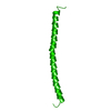 1eq7S  1jcb S: Starting model for refinement C: citing same article ( |
|---|---|
| Similar structure data |


-Links
 PDBj
PDBj


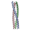
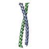
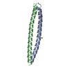
- Assembly
| Deposited unit | 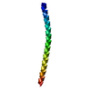
| ||||||||||
|---|---|---|---|---|---|---|---|---|---|---|---|
| 1 | 
| ||||||||||
| Unit cell |
| ||||||||||
| Details | The biological assembly is a trimer generated from the monomer by the three fold axis. |
-Components
| #1: Protein | Mass: 6073.553 Da / Num. of mol.: 1 / Mutation: M30A, V34A Source method: isolated from a genetically manipulated source Source: (gene. exp.) |
|---|---|
| #2: Water | ChemComp-HOH /  Mass: 18.015 Da / Num. of mol.: 19 / Source method: isolated from a natural source / Formula: H2O Mass: 18.015 Da / Num. of mol.: 19 / Source method: isolated from a natural source / Formula: H2O |
-Experimental details
-Experiment
| Experiment | Method: X-RAY DIFFRACTION / Number of used crystals: 1 |
|---|
- Sample preparation
| Crystal | Density Matthews: 1.78 Å3/Da / Density % sol: 30.98 % | ||||||||||||||||||||||||||||||
|---|---|---|---|---|---|---|---|---|---|---|---|---|---|---|---|---|---|---|---|---|---|---|---|---|---|---|---|---|---|---|---|
| Crystal grow | Temperature: 293 K / Method: vapor diffusion, hanging drop / pH: 6.8 Details: PEG 4000, sodium cacodylate, ammonium acetate, pH 6.8, VAPOR DIFFUSION, HANGING DROP at 293K, VAPOR DIFFUSION, HANGING DROP | ||||||||||||||||||||||||||||||
| Crystal grow | *PLUS pH: 7 | ||||||||||||||||||||||||||||||
| Components of the solutions | *PLUS
|
-Data collection
| Diffraction | Mean temperature: 95 K |
|---|---|
| Diffraction source | Source: SYNCHROTRON / Site: NSLS  / Beamline: X25 / Wavelength: 1.1 Å / Beamline: X25 / Wavelength: 1.1 Å |
| Detector | Type: BRANDEIS - B4 / Detector: CCD / Date: Oct 12, 2001 |
| Radiation | Monochromator: GRAPHITE / Protocol: SINGLE WAVELENGTH / Monochromatic (M) / Laue (L): M / Scattering type: x-ray |
| Radiation wavelength | Wavelength: 1.1 Å / Relative weight: 1 |
| Reflection | Resolution: 2→50 Å / Num. all: 2761 / Num. obs: 2761 / % possible obs: 97.8 % / Observed criterion σ(F): 0 / Observed criterion σ(I): 0 / Redundancy: 3.8 % / Biso Wilson estimate: 39 Å2 / Rmerge(I) obs: 0.042 / Net I/σ(I): 17.5 |
| Reflection shell | Resolution: 2→2.07 Å / Redundancy: 3.9 % / Rmerge(I) obs: 0.097 / Mean I/σ(I) obs: 12.3 / Num. unique all: 267 / % possible all: 97 |
| Reflection | *PLUS Highest resolution: 2 Å / Lowest resolution: 50 Å / Num. measured all: 20868 / Rmerge(I) obs: 0.042 |
| Reflection shell | *PLUS Highest resolution: 2 Å / Rmerge(I) obs: 0.097 |
- Processing
| Software |
| ||||||||||||||||||||||||||||||||||||||||||||||||||||||||||||||||||||||||||||||||
|---|---|---|---|---|---|---|---|---|---|---|---|---|---|---|---|---|---|---|---|---|---|---|---|---|---|---|---|---|---|---|---|---|---|---|---|---|---|---|---|---|---|---|---|---|---|---|---|---|---|---|---|---|---|---|---|---|---|---|---|---|---|---|---|---|---|---|---|---|---|---|---|---|---|---|---|---|---|---|---|---|---|
| Refinement | Method to determine structure: MOLECULAR REPLACEMENT Starting model: PDB ENTRY 1EQ7 Resolution: 2→25.26 Å / Rfactor Rfree error: 0.018 / Isotropic thermal model: RESTRAINED / Cross valid method: THROUGHOUT / σ(F): 0 / σ(I): 0 / Stereochemistry target values: Engh & Huber
| ||||||||||||||||||||||||||||||||||||||||||||||||||||||||||||||||||||||||||||||||
| Solvent computation | Solvent model: FLAT MODEL / Bsol: 62.1498 Å2 / ksol: 0.425897 e/Å3 | ||||||||||||||||||||||||||||||||||||||||||||||||||||||||||||||||||||||||||||||||
| Displacement parameters | Biso mean: 43.6 Å2
| ||||||||||||||||||||||||||||||||||||||||||||||||||||||||||||||||||||||||||||||||
| Refine analyze |
| ||||||||||||||||||||||||||||||||||||||||||||||||||||||||||||||||||||||||||||||||
| Refinement step | Cycle: LAST / Resolution: 2→25.26 Å
| ||||||||||||||||||||||||||||||||||||||||||||||||||||||||||||||||||||||||||||||||
| Refine LS restraints |
| ||||||||||||||||||||||||||||||||||||||||||||||||||||||||||||||||||||||||||||||||
| LS refinement shell | Resolution: 2→2.07 Å / Rfactor Rfree error: 0.074 / Total num. of bins used: 10
| ||||||||||||||||||||||||||||||||||||||||||||||||||||||||||||||||||||||||||||||||
| Xplor file |
| ||||||||||||||||||||||||||||||||||||||||||||||||||||||||||||||||||||||||||||||||
| Refinement | *PLUS Highest resolution: 2 Å / Lowest resolution: 50 Å / % reflection Rfree: 10 % / Rfactor obs: 0.26 / Rfactor Rfree: 0.288 / Rfactor Rwork: 0.26 | ||||||||||||||||||||||||||||||||||||||||||||||||||||||||||||||||||||||||||||||||
| Solvent computation | *PLUS | ||||||||||||||||||||||||||||||||||||||||||||||||||||||||||||||||||||||||||||||||
| Displacement parameters | *PLUS | ||||||||||||||||||||||||||||||||||||||||||||||||||||||||||||||||||||||||||||||||
| Refine LS restraints | *PLUS
| ||||||||||||||||||||||||||||||||||||||||||||||||||||||||||||||||||||||||||||||||
| LS refinement shell | *PLUS Highest resolution: 2 Å / Rfactor Rfree: 0.356 / Rfactor Rwork: 0.353 |
