 Movie
Movie Controller

[English] 日本語
 Yorodumi
- PDB-1kfi: Crystal Structure of the Exocytosis-Sensitive Phosphoprotein, pp6... -
+ Open data

- Basic information
| Entry | Database: PDB / ID: 1kfi | ||||||
|---|---|---|---|---|---|---|---|
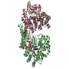
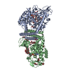
| Title | Crystal Structure of the Exocytosis-Sensitive Phosphoprotein, pp63/Parafusin (phosphoglucomutase) from Paramecium | ||||||
 Components Components | phosphoglucomutase 1 | ||||||
 Keywords Keywords | ISOMERASE / parafusin / phosphoprotein pp63 / exocytosis | ||||||
| Function / homology |  Function and homology information Function and homology informationphosphoglucomutase (alpha-D-glucose-1,6-bisphosphate-dependent) / phosphoglucomutase activity / carbohydrate metabolic process / magnesium ion binding / cytosol Similarity search - Function | ||||||
| Biological species |  | ||||||
| Method |  X-RAY DIFFRACTION / SYNCHROTRON / MOLECULAR REPLACEMENT / Resolution: 2.4 Å X-RAY DIFFRACTION / SYNCHROTRON / MOLECULAR REPLACEMENT / Resolution: 2.4 Å | ||||||
 Authors Authors | Mueller, S. / Diederichs, K. / Breed, J. / Kissmehl, R. / Hauser, K. / Plattner, H. / Welte, W. | ||||||
 Citation Citation | Journal: J.Mol.Biol. / Year: 2002 Title: Crystal structure analysis of the exocytosis-sensitive phosphoprotein, pp63/parafusin (phosphoglucomutase), from Paramecium reveals significant conformational variability. Authors: Muller, S. / Diederichs, K. / Breed, J. / Kissmehl, R. / Hauser, K. / Plattner, H. / Welte, W. | ||||||
| History |
|
- Structure visualization
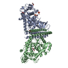
| Structure viewer | Molecule: MolmilJmol/JSmol |
|---|
- Downloads & links
-Download
| PDBx/mmCIF format | 1kfi.cif.gz | 232 KB | Display | PDBx/mmCIF format |
|---|---|---|---|---|
| PDB format | pdb1kfi.ent.gz | 188.3 KB | Display | PDB format |
| PDBx/mmJSON format | 1kfi.json.gz | Tree view | PDBx/mmJSON format | |
| Others |  Other downloads Other downloads |
-Validation report
| Summary document | 1kfi_validation.pdf.gz | 453.4 KB | Display | wwPDB validaton report |
|---|---|---|---|---|
| Full document | 1kfi_full_validation.pdf.gz | 493.4 KB | Display | |
| Data in XML | 1kfi_validation.xml.gz | 46.2 KB | Display | |
| Data in CIF | 1kfi_validation.cif.gz | 63 KB | Display | |
| Arichive directory | https://data.pdbj.org/pub/pdb/validation_reports/kf/1kfiftp://data.pdbj.org/pub/pdb/validation_reports/kf/1kfi | HTTPS FTP |
-Related structure data

-Links
 PDBj
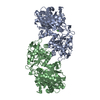
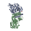
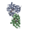
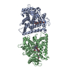
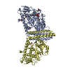
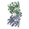
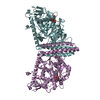
PDBj- Assembly
| Deposited unit | 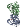
| ||||||||
|---|---|---|---|---|---|---|---|---|---|
| 1 |
| ||||||||
| Unit cell |
|
-Components
| #1: Protein | Mass: 63881.820 Da / Num. of mol.: 2 Source method: isolated from a genetically manipulated source Source: (gene. exp.)  References: UniProt: P47244, phosphoglucomutase (alpha-D-glucose-1,6-bisphosphate-dependent) #2: Chemical |   Mass: 65.409 Da / Num. of mol.: 2 / Source method: obtained synthetically / Formula: Zn Mass: 65.409 Da / Num. of mol.: 2 / Source method: obtained synthetically / Formula: Zn#3: Chemical | ChemComp-SO4 /   Mass: 96.063 Da / Num. of mol.: 4 / Source method: obtained synthetically / Formula: SO4 Mass: 96.063 Da / Num. of mol.: 4 / Source method: obtained synthetically / Formula: SO4#4: Water | ChemComp-HOH / |  Mass: 18.015 Da / Num. of mol.: 205 / Source method: isolated from a natural source / Formula: H2O Mass: 18.015 Da / Num. of mol.: 205 / Source method: isolated from a natural source / Formula: H2O |
|---|
-Experimental details
-Experiment
| Experiment | Method: X-RAY DIFFRACTION / Number of used crystals: 1 |
|---|
- Sample preparation
| Crystal | Density Matthews: 2.65 Å3/Da / Density % sol: 53.58 % | ||||||||||||||||||||||||||||||||||||
|---|---|---|---|---|---|---|---|---|---|---|---|---|---|---|---|---|---|---|---|---|---|---|---|---|---|---|---|---|---|---|---|---|---|---|---|---|---|
| Crystal grow | Temperature: 290 K / Method: vapor diffusion, hanging drop / pH: 4.6 Details: 18 %PEG-MME 2000, 0.2M ammonium sulfate, 0.1M sodium acetate, pH 4.6, VAPOR DIFFUSION, HANGING DROP, temperature 290K | ||||||||||||||||||||||||||||||||||||
| Crystal grow | *PLUS pH: 7.5 | ||||||||||||||||||||||||||||||||||||
| Components of the solutions | *PLUS
|
-Data collection
| Diffraction | Mean temperature: 100 K |
|---|---|
| Diffraction source | Source: SYNCHROTRON / Site: EMBL/DESY, HAMBURG  / Beamline: BW7B / Wavelength: 0.8345 Å / Beamline: BW7B / Wavelength: 0.8345 Å |
| Detector | Type: MARRESEARCH / Detector: IMAGE PLATE / Date: Aug 18, 1998 |
| Radiation | Protocol: SINGLE WAVELENGTH / Monochromatic (M) / Laue (L): M / Scattering type: x-ray |
| Radiation wavelength | Wavelength: 0.8345 Å / Relative weight: 1 |
| Reflection | Resolution: 2.4→30 Å / Num. obs: 50610 / % possible obs: 94.2 % / Observed criterion σ(F): 0 / Observed criterion σ(I): 0 / Redundancy: 3.6 % |
| Reflection shell | Resolution: 2.4→2.49 Å / Redundancy: 2.7 % / Num. unique all: 4304 / % possible all: 77.6 |
| Reflection | *PLUS Lowest resolution: 30 Å / Num. measured all: 180300 / Rmerge(I) obs: 0.041 |
| Reflection shell | *PLUS Highest resolution: 2.4 Å / % possible obs: 77.6 % / Num. unique obs: 4304 / Num. measured obs: 11572 / Rmerge(I) obs: 0.085 / Mean I/σ(I) obs: 11.9 |
- Processing
| Software |
| ||||||||||||||||
|---|---|---|---|---|---|---|---|---|---|---|---|---|---|---|---|---|---|
| Refinement | Method to determine structure: MOLECULAR REPLACEMENT / Resolution: 2.4→30 Å / σ(F): 2.4 / Stereochemistry target values: Engh & Huber
| ||||||||||||||||
| Refinement step | Cycle: LAST / Resolution: 2.4→30 Å
| ||||||||||||||||
| Refine LS restraints | Type: c_bond_d / Dev ideal: 0.007 | ||||||||||||||||
| Software | *PLUS Name: CNS / Classification: refinement | ||||||||||||||||
| Refinement | *PLUS Highest resolution: 2.4 Å / Lowest resolution: 30 Å / σ(F): 2.4 / Rfactor obs: 0.233 / Rfactor Rfree: 0.284 | ||||||||||||||||
| Solvent computation | *PLUS | ||||||||||||||||
| Displacement parameters | *PLUS | ||||||||||||||||
| Refine LS restraints | *PLUS
|
