 Movie
Movie Controller

[English] 日本語
 Yorodumi
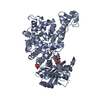
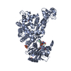
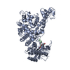
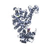
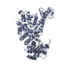
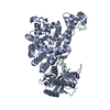
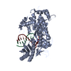
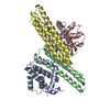
- PDB-1kfd: CRYSTAL STRUCTURES OF THE KLENOW FRAGMENT OF DNA POLYMERASE I COM... -
+ Open data

- Basic information
| Entry | Database: PDB / ID: 1kfd | ||||||
|---|---|---|---|---|---|---|---|
| Title | CRYSTAL STRUCTURES OF THE KLENOW FRAGMENT OF DNA POLYMERASE I COMPLEXED WITH DEOXYNUCLEOSIDE TRIPHOSPHATE AND PYROPHOSPHATE | ||||||
 Components Components | DNA POLYMERASE I KLENOW FRAGMENT | ||||||
 Keywords Keywords | NUCLEOTIDYLTRANSFERASE | ||||||
| Function / homology |  Function and homology information Function and homology information5'-3' exonuclease activity / 3'-5' exonuclease activity / base-excision repair / DNA-templated DNA replication / double-strand break repair / DNA-directed DNA polymerase / DNA-directed DNA polymerase activity / DNA replication / DNA repair / DNA binding ...5'-3' exonuclease activity / 3'-5' exonuclease activity / base-excision repair / DNA-templated DNA replication / double-strand break repair / DNA-directed DNA polymerase / DNA-directed DNA polymerase activity / DNA replication / DNA repair / DNA binding / cytoplasm / cytosol Similarity search - Function | ||||||
| Biological species |  | ||||||
| Method |  X-RAY DIFFRACTION / Resolution: 3.9 Å X-RAY DIFFRACTION / Resolution: 3.9 Å | ||||||
 Authors Authors | Beese, L.S. / Friedman, J.M. / Steitz, T.A. | ||||||
 Citation Citation | Journal: Biochemistry / Year: 1993 Title: Crystal structures of the Klenow fragment of DNA polymerase I complexed with deoxynucleoside triphosphate and pyrophosphate. Authors: Beese, L.S. / Friedman, J.M. / Steitz, T.A. #1: Journal: Science / Year: 1993Title: Structure of DNA Polymerase I Klenow Fragment Bound to Duplex DNA Authors: Beese, L.S. / Derbyshire, V. / Steitz, T.A. #2: Journal: Embo J. / Year: 1991Title: Structural Basis for the 3'-5' Exonuclease Activity of E. Coli DNA Polymerase F: A Two Metal Ion Mechanism Authors: Beese, L.S. / Steitz, T.A. #3: Journal: Proc.Natl.Acad.Sci.USA / Year: 1988Title: Co-Crystal Structure of an Editing Complex of Klenow Fragment with DNA Authors: Freemont, P.S. / Friedman, J.M. / Beese, L.S. / Steitz, T.A. #4: Journal: Nature / Year: 1985Title: Structure of Large Fragment of Escherichia Coli DNA Polymerase I Complexed with dTMP Authors: Ollis, D.L. / Brick, P. / Hamlin, R. / Xuong, N.G. / Steitz, T.A. | ||||||
| History |
|
- Structure visualization
| Structure viewer | Molecule: MolmilJmol/JSmol |
|---|
- Downloads & links
-Download
| PDBx/mmCIF format | 1kfd.cif.gz | 110.5 KB | Display | PDBx/mmCIF format |
|---|---|---|---|---|
| PDB format | pdb1kfd.ent.gz | 82 KB | Display | PDB format |
| PDBx/mmJSON format | 1kfd.json.gz | Tree view | PDBx/mmJSON format | |
| Others |  Other downloads Other downloads |
-Validation report
| Arichive directory | https://data.pdbj.org/pub/pdb/validation_reports/kf/1kfdftp://data.pdbj.org/pub/pdb/validation_reports/kf/1kfd | HTTPS FTP |
|---|
-Related structure data
| Similar structure data |
|---|


-Links
 PDBj
PDBj


- Assembly
| Deposited unit | 
| ||||||||
|---|---|---|---|---|---|---|---|---|---|
| 1 |
| ||||||||
| Unit cell |
|
-Components
| #1: Protein | Mass: 68161.688 Da / Num. of mol.: 1 Source method: isolated from a genetically manipulated source Source: (gene. exp.) |
|---|---|
| #2: Chemical | ChemComp-CTP /   Mass: 483.156 Da / Num. of mol.: 1 / Source method: obtained synthetically / Formula: C9H16N3O14P3 Mass: 483.156 Da / Num. of mol.: 1 / Source method: obtained synthetically / Formula: C9H16N3O14P3 |
-Experimental details
-Experiment
| Experiment | Method: X-RAY DIFFRACTION |
|---|
- Sample preparation
| Crystal | Density Matthews: 3.43 Å3/Da / Density % sol: 64.14 % | |||||||||||||||
|---|---|---|---|---|---|---|---|---|---|---|---|---|---|---|---|---|
| Crystal grow | *PLUS Temperature: 18 ℃ / pH: 6 / Method: vapor diffusion / Details: Brick, P., (1983) J.Mol.Biol., 166, 453. | |||||||||||||||
| Components of the solutions | *PLUS
|
-Data collection
| Radiation | Scattering type: x-ray |
|---|---|
| Radiation wavelength | Relative weight: 1 |
- Processing
| Refinement | Highest resolution: 3.9 Å | ||||||||||||
|---|---|---|---|---|---|---|---|---|---|---|---|---|---|
| Refinement step | Cycle: LAST / Highest resolution: 3.9 Å
|
