 Movie
Movie Controller

[English] 日本語
 Yorodumi
- PDB-1qsl: KLENOW FRAGMENT COMPLEXED WITH SINGLE-STRANDED SUBSTRATE AND EURO... -
+ Open data

- Basic information
| Entry | Database: PDB / ID: 1qsl | ||||||
|---|---|---|---|---|---|---|---|
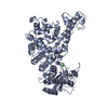
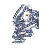
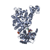
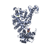
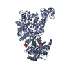
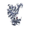
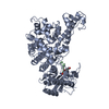
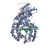
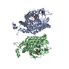
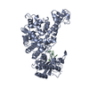
| Title | KLENOW FRAGMENT COMPLEXED WITH SINGLE-STRANDED SUBSTRATE AND EUROPIUM (III) ION | ||||||
 Components Components |
| ||||||
 Keywords Keywords | TRANSFERASE/DNA / EXONUCLEASE / TWO METAL IONS / SINGLE-STRANDED DNA / TRANSFERASE-DNA complex | ||||||
| Function / homology |  Function and homology information Function and homology information5'-3' exonuclease activity / 3'-5' exonuclease activity / base-excision repair / DNA-templated DNA replication / double-strand break repair / DNA-directed DNA polymerase / DNA-directed DNA polymerase activity / DNA replication / DNA repair / DNA binding ...5'-3' exonuclease activity / 3'-5' exonuclease activity / base-excision repair / DNA-templated DNA replication / double-strand break repair / DNA-directed DNA polymerase / DNA-directed DNA polymerase activity / DNA replication / DNA repair / DNA binding / cytosol / cytoplasm Similarity search - Function | ||||||
| Biological species |  | ||||||
| Method |  X-RAY DIFFRACTION / SYNCHROTRON / Resolution: 2.2 Å X-RAY DIFFRACTION / SYNCHROTRON / Resolution: 2.2 Å | ||||||
 Authors Authors | Brautigam, C.A. / Aschheim, K. / Steitz, T.A. | ||||||
 Citation Citation | Journal: Chem.Biol. / Year: 1999 Title: Structural elucidation of the binding and inhibitory properties of lanthanide (III) ions at the 3'-5' exonucleolytic active site of the Klenow fragment Authors: Brautigam, C.A. / Aschheim, K. / Steitz, T.A. #1: Journal: J.Mol.Biol. / Year: 1998Title: Structural Principles for the Inhibition of the 3'-5' Exonuclease Activity of Escherichia Coli DNA Polymerase I by Phosphorothioates Authors: Brautigam, C.A. / Steitz, T.A. #2: Journal: Embo J. / Year: 1991Title: Structural Basis for the 3'-5' Exonuclease Activity of Escherichia Coli DNA Polymerase I: A Two Metal Ion Mechanism Authors: Beese, L.S. / Steitz, T.A. | ||||||
| History |
|
- Structure visualization
| Structure viewer | Molecule: MolmilJmol/JSmol |
|---|
- Downloads & links
-Download
| PDBx/mmCIF format | 1qsl.cif.gz | 140.5 KB | Display | PDBx/mmCIF format |
|---|---|---|---|---|
| PDB format | pdb1qsl.ent.gz | 106.4 KB | Display | PDB format |
| PDBx/mmJSON format | 1qsl.json.gz | Tree view | PDBx/mmJSON format | |
| Others |  Other downloads Other downloads |
-Validation report
| Arichive directory | https://data.pdbj.org/pub/pdb/validation_reports/qs/1qslftp://data.pdbj.org/pub/pdb/validation_reports/qs/1qsl | HTTPS FTP |
|---|
-Related structure data
| Similar structure data |
|---|
-Links
 PDBj
PDBj


- Assembly
| Deposited unit | 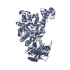
| ||||||||||
|---|---|---|---|---|---|---|---|---|---|---|---|
| 1 |
| ||||||||||
| Unit cell |
|
-Components
| #1: DNA chain | Mass: 2402.592 Da / Num. of mol.: 1 / Source method: obtained synthetically Details: CHEMICALLY SYNTHESIZED BY STANDARD PHOSPHORAMIDITE METHODOLOGY |
|---|---|
| #2: Protein | Mass: 68193.750 Da / Num. of mol.: 1 / Fragment: KLENOW FRAGMENT / Mutation: V324M Source method: isolated from a genetically manipulated source Source: (gene. exp.) |
| #3: Chemical | ChemComp-EU /   Mass: 151.964 Da / Num. of mol.: 1 / Source method: obtained synthetically / Formula: Eu Mass: 151.964 Da / Num. of mol.: 1 / Source method: obtained synthetically / Formula: Eu |
| #4: Water | ChemComp-HOH /  Mass: 18.015 Da / Num. of mol.: 191 / Source method: isolated from a natural source / Formula: H2O Mass: 18.015 Da / Num. of mol.: 191 / Source method: isolated from a natural source / Formula: H2O |
-Experimental details
-Experiment
| Experiment | Method: X-RAY DIFFRACTION / Number of used crystals: 1 |
|---|
- Sample preparation
| Crystal | Density Matthews: 3.26 Å3/Da / Density % sol: 62.3 % | |||||||||||||||
|---|---|---|---|---|---|---|---|---|---|---|---|---|---|---|---|---|
| Crystal grow | Temperature: 290 K / Method: vapor diffusion, hanging drop / pH: 5.6 Details: 1.5 M SODIUM CITRATE AT 290 K, pH 5.60, VAPOR DIFFUSION, HANGING DROP | |||||||||||||||
| Components of the solutions |
| |||||||||||||||
| Crystal grow | *PLUS Temperature: 18 ℃ / pH: 6 / Method: vapor diffusion | |||||||||||||||
| Components of the solutions | *PLUS
|
-Data collection
| Diffraction | Mean temperature: 100 K |
|---|---|
| Diffraction source | Source: SYNCHROTRON / Site: CHESS  / Beamline: A1 / Wavelength: 0.9 / Beamline: A1 / Wavelength: 0.9 |
| Detector | Type: ADSC / Detector: CCD / Date: May 17, 1997 |
| Radiation | Protocol: SINGLE WAVELENGTH / Monochromatic (M) / Laue (L): M / Scattering type: x-ray |
| Radiation wavelength | Wavelength: 0.9 Å / Relative weight: 1 |
| Reflection | Resolution: 2.2→20 Å / Num. obs: 45999 / % possible obs: 99.6 % / Observed criterion σ(I): -3 / Redundancy: 3.7 % / Biso Wilson estimate: 38 Å2 / Rmerge(I) obs: 0.074 / Net I/σ(I): 16.4 |
| Reflection shell | Resolution: 2.2→2.23 Å / Redundancy: 3.5 % / Rmerge(I) obs: 0.346 / % possible all: 99.7 |
| Reflection | *PLUS Num. obs: 40431 / Num. measured all: 343566 |
| Reflection shell | *PLUS Rmerge(I) obs: 0.345 / Mean I/σ(I) obs: 3.7 |
- Processing
| Software |
| ||||||||||||||||||||||||||||||||||||||||||||||||||||||||||||
|---|---|---|---|---|---|---|---|---|---|---|---|---|---|---|---|---|---|---|---|---|---|---|---|---|---|---|---|---|---|---|---|---|---|---|---|---|---|---|---|---|---|---|---|---|---|---|---|---|---|---|---|---|---|---|---|---|---|---|---|---|---|
| Refinement | Resolution: 2.2→20 Å / σ(F): 2 / Details: BULK-SOLVENT CORRECTION WAS APPLIED
| ||||||||||||||||||||||||||||||||||||||||||||||||||||||||||||
| Refinement step | Cycle: LAST / Resolution: 2.2→20 Å
| ||||||||||||||||||||||||||||||||||||||||||||||||||||||||||||
| Refine LS restraints |
| ||||||||||||||||||||||||||||||||||||||||||||||||||||||||||||
| Software | *PLUS Name: X-PLOR / Version: 3.851 / Classification: refinement | ||||||||||||||||||||||||||||||||||||||||||||||||||||||||||||
| Refine LS restraints | *PLUS
|
