 Movie
Movie Controller

[English] 日本語
 Yorodumi
- PDB-1keq: Crystal Structure of F65A/Y131C Carbonic Anhydrase V, covalently ... -
+ Open data

- Basic information
| Entry | Database: PDB / ID: 1keq | ||||||
|---|---|---|---|---|---|---|---|
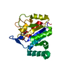
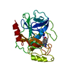
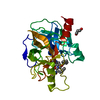
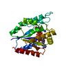
| Title | Crystal Structure of F65A/Y131C Carbonic Anhydrase V, covalently modified with 4-chloromethylimidazole | ||||||
 Components Components | F65A/Y131C-MI Carbonic Anhydrase V | ||||||
 Keywords Keywords | LYASE / PROTON TRANSFER / ENGINEERED RESIDUE | ||||||
| Function / homology |  Function and homology information Function and homology informationReversible hydration of carbon dioxide / carbonic anhydrase / gluconeogenesis / carbonate dehydratase activity / mitochondrion / zinc ion binding / cytoplasm Similarity search - Function | ||||||
| Biological species |  | ||||||
| Method |  X-RAY DIFFRACTION / SYNCHROTRON / MOLECULAR REPLACEMENT / Resolution: 1.88 Å X-RAY DIFFRACTION / SYNCHROTRON / MOLECULAR REPLACEMENT / Resolution: 1.88 Å | ||||||
 Authors Authors | Jude, K.M. / Wright, S.K. / Tu, C. / Silverman, D.N. / Viola, R.E. / Christianson, D.W. | ||||||
 Citation Citation | Journal: Biochemistry / Year: 2002 Title: Crystal structure of F65A/Y131C-methylimidazole carbonic anhydrase V reveals architectural features of an engineered proton shuttle. Authors: Jude, K.M. / Wright, S.K. / Tu, C. / Silverman, D.N. / Viola, R.E. / Christianson, D.W. #1: Journal: Arch.Biochem.Biophys. / Year: 1999Title: Introduction of Histidine Analogs Leads to Enhanced Proton Transfer in Carbonic Anhydrase V Authors: Earnhardt, J.N. / Wright, S.K. / Qian, M. / Tu, C. / Laipis, P.J. / Viola, R.E. / Silverman, D.N. #2: Journal: Proc.Natl.Acad.Sci.USA / Year: 1995Title: Structure determination of murine mitochondrial carbonic anhydrase V at 2.45-A resolution: implications for catalytic proton transfer and inhibitor design. Authors: Boriack-Sjodin, P.A. / Heck, R.W. / Laipis, P.J. / Silverman, D.N. / Christianson, D.W. | ||||||
| History |
|
- Structure visualization
| Structure viewer | Molecule: MolmilJmol/JSmol |
|---|
- Downloads & links
-Download
| PDBx/mmCIF format | 1keq.cif.gz | 120.5 KB | Display | PDBx/mmCIF format |
|---|---|---|---|---|
| PDB format | pdb1keq.ent.gz | 92 KB | Display | PDB format |
| PDBx/mmJSON format | 1keq.json.gz | Tree view | PDBx/mmJSON format | |
| Others |  Other downloads Other downloads |
-Validation report
| Arichive directory | https://data.pdbj.org/pub/pdb/validation_reports/ke/1keqftp://data.pdbj.org/pub/pdb/validation_reports/ke/1keq | HTTPS FTP |
|---|
-Related structure data
| Related structure data |  1dmxS S: Starting model for refinement |
|---|---|
| Similar structure data |


-Links
 PDBj
PDBj

- Assembly
| Deposited unit | 
| ||||||||
|---|---|---|---|---|---|---|---|---|---|
| 1 | 
| ||||||||
| 2 | 
| ||||||||
| Unit cell |
|
-Components
-Protein , 1 types, 2 molecules AB
| #1: Protein | Mass: 28139.725 Da / Num. of mol.: 2 / Fragment: Carbonic Anhydrase Vc Mutation: Truncation of mitochondrial leader sequence and first 21 residues, F65A/Y131C Source method: isolated from a genetically manipulated source Source: (gene. exp.)  |
|---|
-Non-polymers , 5 types, 482 molecules 


| #2: Chemical | ChemComp-K /  Mass: 39.098 Da / Num. of mol.: 1 / Source method: obtained synthetically / Formula: K Mass: 39.098 Da / Num. of mol.: 1 / Source method: obtained synthetically / Formula: K | ||||||
|---|---|---|---|---|---|---|---|
| #3: Chemical |  Mass: 65.409 Da / Num. of mol.: 2 / Source method: obtained synthetically / Formula: Zn Mass: 65.409 Da / Num. of mol.: 2 / Source method: obtained synthetically / Formula: Zn#4: Chemical | ChemComp-4MZ /  Mass: 82.104 Da / Num. of mol.: 5 / Source method: obtained synthetically / Formula: C4H6N2 Mass: 82.104 Da / Num. of mol.: 5 / Source method: obtained synthetically / Formula: C4H6N2#5: Chemical |  Mass: 60.052 Da / Num. of mol.: 3 / Source method: obtained synthetically / Formula: C2H4O2 Mass: 60.052 Da / Num. of mol.: 3 / Source method: obtained synthetically / Formula: C2H4O2#6: Water | ChemComp-HOH / | Mass: 18.015 Da / Num. of mol.: 471 / Source method: isolated from a natural source / Formula: H2O |
-Details
| Has protein modification | Y |
|---|
-Experimental details
-Experiment
| Experiment | Method: X-RAY DIFFRACTION / Number of used crystals: 1 |
|---|
- Sample preparation
| Crystal | Density Matthews: 2.61 Å3/Da / Density % sol: 52.45 % | ||||||||||||||||||||||||||||||||||||||||||
|---|---|---|---|---|---|---|---|---|---|---|---|---|---|---|---|---|---|---|---|---|---|---|---|---|---|---|---|---|---|---|---|---|---|---|---|---|---|---|---|---|---|---|---|
| Crystal grow | Temperature: 293 K / Method: vapor diffusion, hanging drop / pH: 6.7 Details: PEG 8000, sodium acetate, DTT, sucrose monolaurate, MES, pH 6.7, VAPOR DIFFUSION, HANGING DROP, temperature 293K | ||||||||||||||||||||||||||||||||||||||||||
| Crystal grow | *PLUS | ||||||||||||||||||||||||||||||||||||||||||
| Components of the solutions | *PLUS
|
-Data collection
| Diffraction | Mean temperature: 100 K |
|---|---|
| Diffraction source | Source: SYNCHROTRON / Site: NSLS  / Beamline: X12B / Wavelength: 0.9796 Å / Beamline: X12B / Wavelength: 0.9796 Å |
| Detector | Type: ADSC QUANTUM 4 / Detector: CCD / Date: Apr 17, 2001 / Details: mirrors |
| Radiation | Monochromator: Double-crystal, fixed-exit Si-III / Protocol: SINGLE WAVELENGTH / Monochromatic (M) / Laue (L): M / Scattering type: x-ray |
| Radiation wavelength | Wavelength: 0.9796 Å / Relative weight: 1 |
| Reflection | Resolution: 1.85→25 Å / Num. obs: 46338 / % possible obs: 97.6 % / Observed criterion σ(I): -3 / Redundancy: 3 % / Biso Wilson estimate: 16.5 Å2 / Rmerge(I) obs: 0.051 / Net I/σ(I): 13.1 |
| Reflection shell | Resolution: 1.85→1.92 Å / Redundancy: 3 % / Rmerge(I) obs: 0.217 / Mean I/σ(I) obs: 3 / Num. unique all: 4557 / % possible all: 96.4 |
| Reflection | *PLUS Highest resolution: 1.88 Å / Lowest resolution: 25 Å / Num. obs: 46312 / % possible obs: 96.8 % / Num. measured all: 141410 |
| Reflection shell | *PLUS % possible obs: 87.9 % |
- Processing
| Software |
| ||||||||||||||||||||||||||||||||||||||||
|---|---|---|---|---|---|---|---|---|---|---|---|---|---|---|---|---|---|---|---|---|---|---|---|---|---|---|---|---|---|---|---|---|---|---|---|---|---|---|---|---|---|
| Refinement | Method to determine structure: MOLECULAR REPLACEMENT Starting model: PDB ENTRY 1DMX Resolution: 1.88→24.56 Å / Rfactor Rfree error: 0.005 / Data cutoff high absF: 253283.34 / Data cutoff low absF: 0 / Isotropic thermal model: RESTRAINED / Cross valid method: THROUGHOUT / σ(F): 0 Stereochemistry target values: Examples of Stereochemistry target values (mmCIF item _refine.ndb_stereochemistry_target_values) Example 1: Engh & Huber Details: Maximum likelihood
| ||||||||||||||||||||||||||||||||||||||||
| Solvent computation | Solvent model: FLAT MODEL / Bsol: 50.0435 Å2 / ksol: 0.362292 e/Å3 | ||||||||||||||||||||||||||||||||||||||||
| Displacement parameters | Biso mean: 18 Å2
| ||||||||||||||||||||||||||||||||||||||||
| Refine analyze |
| ||||||||||||||||||||||||||||||||||||||||
| Refinement step | Cycle: LAST / Resolution: 1.88→24.56 Å
| ||||||||||||||||||||||||||||||||||||||||
| Refine LS restraints |
| ||||||||||||||||||||||||||||||||||||||||
| LS refinement shell | Resolution: 1.88→1.99 Å / Rfactor Rfree error: 0.014 / Total num. of bins used: 6
| ||||||||||||||||||||||||||||||||||||||||
| Xplor file |
| ||||||||||||||||||||||||||||||||||||||||
| Refinement | *PLUS Lowest resolution: 25 Å / Num. reflection obs: 44468 / σ(F): 0 / % reflection Rfree: 4 % / Rfactor obs: 0.191 / Rfactor Rfree: 0.222 | ||||||||||||||||||||||||||||||||||||||||
| Solvent computation | *PLUS | ||||||||||||||||||||||||||||||||||||||||
| Displacement parameters | *PLUS Biso mean: 18 Å2 | ||||||||||||||||||||||||||||||||||||||||
| Refine LS restraints | *PLUS
| ||||||||||||||||||||||||||||||||||||||||
| LS refinement shell | *PLUS Rfactor Rfree: 0.234 / % reflection Rfree: 3.9 % / Rfactor Rwork: 0.22 |
