 Movie
Movie Controller

[English] 日本語
 Yorodumi
- PDB-1kd8: X-RAY STRUCTURE OF THE COILED COIL GCN4 ACID BASE HETERODIMER ACI... -
+ Open data

- Basic information
| Entry | Database: PDB / ID: 1kd8 | ||||||
|---|---|---|---|---|---|---|---|
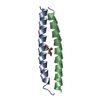
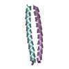
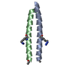
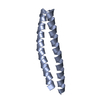
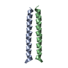
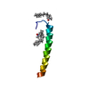
| Title | X-RAY STRUCTURE OF THE COILED COIL GCN4 ACID BASE HETERODIMER ACID-d12Ia16V BASE-d12La16L | ||||||
 Components Components |
| ||||||
 Keywords Keywords | DE NOVO PROTEIN / coiled coil heterodimer | ||||||
| Method |  X-RAY DIFFRACTION / SYNCHROTRON / MOLECULAR REPLACEMENT / Resolution: 1.9 Å X-RAY DIFFRACTION / SYNCHROTRON / MOLECULAR REPLACEMENT / Resolution: 1.9 Å | ||||||
 Authors Authors | Keating, A.E. / Malashkevich, V.N. / Tidor, B. / Kim, P.S. | ||||||
 Citation Citation | Journal: Proc.Natl.Acad.Sci.USA / Year: 2001 Title: Side-chain repacking calculations for predicting structures and stabilities of heterodimeric coiled coils. Authors: Keating, A.E. / Malashkevich, V.N. / Tidor, B. / Kim, P.S. | ||||||
| History |
|
- Structure visualization
| Structure viewer | Molecule:  MolmilJmol/JSmol MolmilJmol/JSmol |
|---|
- Downloads & links
-Download
| PDBx/mmCIF format | 1kd8.cif.gz | 57.5 KB | Display | PDBx/mmCIF format |
|---|---|---|---|---|
| PDB format | pdb1kd8.ent.gz | 44.6 KB | Display | PDB format |
| PDBx/mmJSON format | 1kd8.json.gz | Tree view | PDBx/mmJSON format | |
| Others |  Other downloads Other downloads |
-Validation report
| Summary document | 1kd8_validation.pdf.gz | 467.7 KB | Display | wwPDB validaton report |
|---|---|---|---|---|
| Full document | 1kd8_full_validation.pdf.gz | 470.8 KB | Display | |
| Data in XML | 1kd8_validation.xml.gz | 13.4 KB | Display | |
| Data in CIF | 1kd8_validation.cif.gz | 19.4 KB | Display | |
| Arichive directory | https://data.pdbj.org/pub/pdb/validation_reports/kd/1kd8ftp://data.pdbj.org/pub/pdb/validation_reports/kd/1kd8 | HTTPS FTP |
-Related structure data


-Links
 PDBj
PDBj
- Assembly
| Deposited unit | 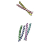
| ||||||||
|---|---|---|---|---|---|---|---|---|---|
| 1 | 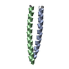
| ||||||||
| 2 | 
| ||||||||
| 3 | 
| ||||||||
| Unit cell |
| ||||||||
| Components on special symmetry positions |
| ||||||||
| Details | The assembly is a dimer. There are three copies of the dimer intact in the asymmetric unit. |
-Components
| #1: Protein/peptide | Mass: 4142.462 Da / Num. of mol.: 3 / Source method: obtained synthetically / Details: The peptide was chemically synthesized. #2: Protein/peptide | Mass: 4157.150 Da / Num. of mol.: 3 / Source method: obtained synthetically / Details: The peptide was chemically synthesized. #3: Water | ChemComp-HOH / |  Mass: 18.015 Da / Num. of mol.: 288 / Source method: isolated from a natural source / Formula: H2O Mass: 18.015 Da / Num. of mol.: 288 / Source method: isolated from a natural source / Formula: H2OHas protein modification | Y | |
|---|
-Experimental details
-Experiment
| Experiment | Method: X-RAY DIFFRACTION / Number of used crystals: 1 |
|---|
- Sample preparation
| Crystal | Density Matthews: 2.91 Å3/Da / Density % sol: 56.1 % | ||||||||||||||||||||||||||||||
|---|---|---|---|---|---|---|---|---|---|---|---|---|---|---|---|---|---|---|---|---|---|---|---|---|---|---|---|---|---|---|---|
| Crystal grow | Temperature: 293 K / Method: vapor diffusion, hanging drop / pH: 7.7 Details: PEG 4000, Na Hepes, 2-propanol, pH 7.7, VAPOR DIFFUSION, HANGING DROP, temperature 293K | ||||||||||||||||||||||||||||||
| Crystal grow | *PLUS | ||||||||||||||||||||||||||||||
| Components of the solutions | *PLUS
|
-Data collection
| Diffraction | Mean temperature: 100 K |
|---|---|
| Diffraction source | Source: SYNCHROTRON / Site: NSLS  / Beamline: X4A / Wavelength: 0.9763 Å / Beamline: X4A / Wavelength: 0.9763 Å |
| Detector | Type: ADSC QUANTUM 4 / Detector: CCD / Date: Oct 19, 2000 |
| Radiation | Protocol: SINGLE WAVELENGTH / Monochromatic (M) / Laue (L): M / Scattering type: x-ray |
| Radiation wavelength | Wavelength: 0.9763 Å / Relative weight: 1 |
| Reflection | Resolution: 1.85→35 Å / Num. all: 25471 / Num. obs: 25471 / % possible obs: 98.9 % / Observed criterion σ(F): 0 / Observed criterion σ(I): 0 / Biso Wilson estimate: 20.8 Å2 / Rmerge(I) obs: 0.06 |
| Reflection shell | Resolution: 1.85→1.92 Å / Rmerge(I) obs: 0.413 / % possible all: 99 |
| Reflection | *PLUS Rmerge(I) obs: 0.06 |
- Processing
| Software |
| |||||||||||||||||||||||||
|---|---|---|---|---|---|---|---|---|---|---|---|---|---|---|---|---|---|---|---|---|---|---|---|---|---|---|
| Refinement | Method to determine structure: MOLECULAR REPLACEMENT Starting model: calculated structure Resolution: 1.9→28.97 Å / Rfactor Rfree error: 0.006 / Isotropic thermal model: restrained / Cross valid method: THROUGHOUT / σ(F): 0 / σ(I): 0 / Stereochemistry target values: Engh & Huber
| |||||||||||||||||||||||||
| Solvent computation | Solvent model: flat model / Bsol: 62.63 Å2 / ksol: 0.386808 e/Å3 | |||||||||||||||||||||||||
| Displacement parameters | Biso mean: 35.8 Å2 | |||||||||||||||||||||||||
| Refine analyze |
| |||||||||||||||||||||||||
| Refinement step | Cycle: LAST / Resolution: 1.9→28.97 Å
| |||||||||||||||||||||||||
| Refine LS restraints |
| |||||||||||||||||||||||||
| LS refinement shell | Resolution: 1.9→2.02 Å / Rfactor Rfree error: 0.017 / Total num. of bins used: 6
| |||||||||||||||||||||||||
| Software | *PLUS Name: CNS / Version: 1 / Classification: refinement | |||||||||||||||||||||||||
| Refinement | *PLUS Lowest resolution: 30 Å / % reflection Rfree: 10 % / Rfactor obs: 0.232 / Rfactor Rfree: 0.276 | |||||||||||||||||||||||||
| Solvent computation | *PLUS | |||||||||||||||||||||||||
| Displacement parameters | *PLUS Biso mean: 35.8 Å2 | |||||||||||||||||||||||||
| Refine LS restraints | *PLUS
| |||||||||||||||||||||||||
| LS refinement shell | *PLUS Rfactor Rfree: 0.328 / % reflection Rfree: 9.9 % / Rfactor Rwork: 0.263 |
