 Movie
Movie Controller

[English] 日本語
 Yorodumi
- PDB-1kcm: Crystal Structure of Mouse PITP Alpha Void of Bound Phospholipid ... -
+ Open data

- Basic information
| Entry | Database: PDB / ID: 1kcm | ||||||
|---|---|---|---|---|---|---|---|
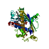
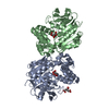
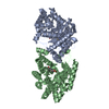
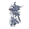
| Title | Crystal Structure of Mouse PITP Alpha Void of Bound Phospholipid at 2.0 Angstroms Resolution | ||||||
 Components Components | Phosphatidylinositol Transfer Protein alpha | ||||||
 Keywords Keywords | LIPID BINDING PROTEIN / PITP / phospholipid binding protein / phospholipid transport | ||||||
| Function / homology |  Function and homology information Function and homology informationstearic acid binding / phosphatidylcholine intramembrane carrier activity / phosphatidylcholine transfer activity / phosphatidylinositol transfer activity / fatty-acyl-CoA binding / phospholipid transport / phosphatidylcholine binding / axonogenesis / phosphatidylinositol binding / phospholipid binding ...stearic acid binding / phosphatidylcholine intramembrane carrier activity / phosphatidylcholine transfer activity / phosphatidylinositol transfer activity / fatty-acyl-CoA binding / phospholipid transport / phosphatidylcholine binding / axonogenesis / phosphatidylinositol binding / phospholipid binding / myelin sheath / lipid binding / membrane / nucleus / cytoplasm / cytosol Similarity search - Function | ||||||
| Biological species |  | ||||||
| Method |  X-RAY DIFFRACTION / SYNCHROTRON / MOLECULAR REPLACEMENT / Resolution: 2 Å X-RAY DIFFRACTION / SYNCHROTRON / MOLECULAR REPLACEMENT / Resolution: 2 Å | ||||||
 Authors Authors | Schouten, A. / Agianian, B. / Westerman, J. / Kroon, J. / Wirtz, K.W.A. / Gros, P. | ||||||
 Citation Citation | Journal: EMBO J. / Year: 2002 Title: Structure of apo-phosphatidylinositol transfer protein alpha provides insight into membrane association. Authors: Schouten, A. / Agianian, B. / Westerman, J. / Kroon, J. / Wirtz, K.W. / Gros, P. | ||||||
| History |
|
- Structure visualization
| Structure viewer | Molecule: MolmilJmol/JSmol |
|---|
- Downloads & links
-Download
| PDBx/mmCIF format | 1kcm.cif.gz | 69.5 KB | Display | PDBx/mmCIF format |
|---|---|---|---|---|
| PDB format | pdb1kcm.ent.gz | 50.8 KB | Display | PDB format |
| PDBx/mmJSON format | 1kcm.json.gz | Tree view | PDBx/mmJSON format | |
| Others |  Other downloads Other downloads |
-Validation report
| Arichive directory | https://data.pdbj.org/pub/pdb/validation_reports/kc/1kcmftp://data.pdbj.org/pub/pdb/validation_reports/kc/1kcm | HTTPS FTP |
|---|
-Related structure data
| Related structure data |  1fvz S: Starting model for refinement |
|---|---|
| Similar structure data |
-Links
 PDBj
PDBj- Assembly
| Deposited unit | 
| ||||||||
|---|---|---|---|---|---|---|---|---|---|
| 1 | 
| ||||||||
| Unit cell |
| ||||||||
| Details | The biological assembly is a homodimer generated from the monomer in the asymmetric unit by the operation: y, x, -z |
-Components
| #1: Protein | Mass: 31810.232 Da / Num. of mol.: 1 Source method: isolated from a genetically manipulated source Source: (gene. exp.) Plasmid details: PET3D CONTAINED PITP cDNA from Swiss mouse 3T3 fibroblast cells Plasmid: pET3D / Species (production host): Escherichia coli / Production host:  |
|---|---|
| #2: Water | ChemComp-HOH /  Mass: 18.015 Da / Num. of mol.: 216 / Source method: isolated from a natural source / Formula: H2O Mass: 18.015 Da / Num. of mol.: 216 / Source method: isolated from a natural source / Formula: H2O |
-Experimental details
-Experiment
| Experiment | Method: X-RAY DIFFRACTION / Number of used crystals: 1 |
|---|
- Sample preparation
| Crystal | Density Matthews: 2.5 Å3/Da / Density % sol: 50.72 % | ||||||||||||||||||||||||||||||||||||||||||
|---|---|---|---|---|---|---|---|---|---|---|---|---|---|---|---|---|---|---|---|---|---|---|---|---|---|---|---|---|---|---|---|---|---|---|---|---|---|---|---|---|---|---|---|
| Crystal grow | Temperature: 277 K / Method: vapor diffusion, hanging drop / pH: 6.5 Details: 11% PEG 6000, 0.2M calcium acetate, 100MM cacodylate, pH 6.5, VAPOR DIFFUSION, HANGING DROP, temperature 277.0K | ||||||||||||||||||||||||||||||||||||||||||
| Crystal grow | *PLUS Temperature: 4 ℃ / pH: 7.2 | ||||||||||||||||||||||||||||||||||||||||||
| Components of the solutions | *PLUS
|
-Data collection
| Diffraction | Mean temperature: 100 K |
|---|---|
| Diffraction source | Source: SYNCHROTRON / Site: ESRF  / Beamline: ID14-4 / Wavelength: 0.9322 Å / Beamline: ID14-4 / Wavelength: 0.9322 Å |
| Detector | Type: ADSC QUANTUM 4r / Detector: CCD / Date: Dec 9, 1999 / Details: Toroidal mirror |
| Radiation | Monochromator: LN2 cooled Si(311) or Si(111) crystal / Protocol: SINGLE WAVELENGTH / Monochromatic (M) / Laue (L): M / Scattering type: x-ray |
| Radiation wavelength | Wavelength: 0.9322 Å / Relative weight: 1 |
| Reflection | Resolution: 1.99→30 Å / Num. all: 22808 / Num. obs: 22060 / % possible obs: 96.7 % / Observed criterion σ(I): -3 / Redundancy: 4.3 % / Biso Wilson estimate: 28.9 Å2 / Rmerge(I) obs: 0.051 / Rsym value: 0.051 / Net I/σ(I): 25.7 |
| Reflection shell | Resolution: 1.99→2.06 Å / Redundancy: 2.7 % / Rmerge(I) obs: 0.171 / Mean I/σ(I) obs: 7.3 / Num. unique all: 2209 / Rsym value: 0.171 / % possible all: 91.1 |
| Reflection | *PLUS Lowest resolution: 30 Å / Redundancy: 4.33 % / Num. measured all: 231585 / Rmerge(I) obs: 0.051 |
| Reflection shell | *PLUS % possible obs: 91.1 % / Redundancy: 2.7 % / Num. unique obs: 2012 / Rmerge(I) obs: 0.171 |
- Processing
| Software |
| ||||||||||||||||||||||||||||||||||||
|---|---|---|---|---|---|---|---|---|---|---|---|---|---|---|---|---|---|---|---|---|---|---|---|---|---|---|---|---|---|---|---|---|---|---|---|---|---|
| Refinement | Method to determine structure: MOLECULAR REPLACEMENT Starting model: PDB ENTRY 1FVZ 1fvz Resolution: 2→30 Å / SU B: 4.23065 / SU ML: 0.12169 / Isotropic thermal model: Isotropic / Cross valid method: THROUGHOUT / σ(F): 0 / ESU R: 0.20144 / ESU R Free: 0.18838 Stereochemistry target values: Engh & Huber, mon_lib (CCP4: Library) Details: Used maximum likelihood refinement, and TLS refinement
| ||||||||||||||||||||||||||||||||||||
| Displacement parameters | Biso mean: 24.34 Å2
| ||||||||||||||||||||||||||||||||||||
| Refine analyze |
| ||||||||||||||||||||||||||||||||||||
| Refinement step | Cycle: LAST / Resolution: 2→30 Å
| ||||||||||||||||||||||||||||||||||||
| Refine LS restraints |
| ||||||||||||||||||||||||||||||||||||
| Refinement | *PLUS Lowest resolution: 30 Å / Rfactor all: 0.2188 / Rfactor obs: 0.216 / Rfactor Rfree: 0.273 / Rfactor Rwork: 0.216 | ||||||||||||||||||||||||||||||||||||
| Solvent computation | *PLUS | ||||||||||||||||||||||||||||||||||||
| Displacement parameters | *PLUS | ||||||||||||||||||||||||||||||||||||
| Refine LS restraints | *PLUS Type: p_angle_d / Dev ideal: 1.42 |
