 Movie
Movie Controller


+ Open data

- Basic information
| Entry | Database: PDB / ID: 1k9i | |||||||||
|---|---|---|---|---|---|---|---|---|---|---|
| Title | Complex of DC-SIGN and GlcNAc2Man3 | |||||||||
 Components Components | mDC-SIGN1B type I isoform | |||||||||
 Keywords Keywords | SUGAR BINDING PROTEIN / C-type lectin / protein carbohydrate complex | |||||||||
| Function / homology |  Function and homology information Function and homology informationB cell adhesion / cell-cell recognition / intracellular transport of virus / peptide antigen transport / Butyrophilin (BTN) family interactions / positive regulation of viral life cycle / virion binding / heterophilic cell-cell adhesion via plasma membrane cell adhesion molecules / leukocyte cell-cell adhesion / pattern recognition receptor activity ...B cell adhesion / cell-cell recognition / intracellular transport of virus / peptide antigen transport / Butyrophilin (BTN) family interactions / positive regulation of viral life cycle / virion binding / heterophilic cell-cell adhesion via plasma membrane cell adhesion molecules / leukocyte cell-cell adhesion / pattern recognition receptor activity / antigen processing and presentation / RSV-host interactions / D-mannose binding / regulation of T cell proliferation / positive regulation of T cell proliferation / CD209 (DC-SIGN) signaling / viral genome replication / peptide antigen binding / endocytosis / host cell / virus receptor activity / carbohydrate binding / adaptive immune response / intracellular signal transduction / immune response / innate immune response / external side of plasma membrane / symbiont entry into host cell / virion attachment to host cell / cell surface / extracellular region / metal ion binding / membrane / plasma membrane / cytoplasm Similarity search - Function | |||||||||
| Biological species |  Homo sapiens (human) Homo sapiens (human) | |||||||||
| Method |  X-RAY DIFFRACTION / SYNCHROTRON / MOLECULAR REPLACEMENT / Resolution: 2.5 Å X-RAY DIFFRACTION / SYNCHROTRON / MOLECULAR REPLACEMENT / Resolution: 2.5 Å | |||||||||
 Authors Authors | Feinberg, H. / Mitchell, D.A. / Drickamer, K. / Weis, W.I. | |||||||||
 Citation Citation | Journal: Science / Year: 2001 Title: Structural basis for selective recognition of oligosaccharides by DC-SIGN and DC-SIGNR. Authors: Feinberg, H. / Mitchell, D.A. / Drickamer, K. / Weis, W.I. | |||||||||
| History |
|
- Structure visualization
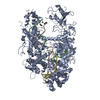
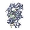
| Structure viewer | Molecule: MolmilJmol/JSmol |
|---|
- Downloads & links
-Download
| PDBx/mmCIF format | 1k9i.cif.gz | 289.8 KB | Display | PDBx/mmCIF format |
|---|---|---|---|---|
| PDB format | pdb1k9i.ent.gz | 232.8 KB | Display | PDB format |
| PDBx/mmJSON format | 1k9i.json.gz | Tree view | PDBx/mmJSON format | |
| Others |  Other downloads Other downloads |
-Validation report
| Summary document | 1k9i_validation.pdf.gz | 2.1 MB | Display | wwPDB validaton report |
|---|---|---|---|---|
| Full document | 1k9i_full_validation.pdf.gz | 2.1 MB | Display | |
| Data in XML | 1k9i_validation.xml.gz | 59.8 KB | Display | |
| Data in CIF | 1k9i_validation.cif.gz | 76.6 KB | Display | |
| Arichive directory | https://data.pdbj.org/pub/pdb/validation_reports/k9/1k9iftp://data.pdbj.org/pub/pdb/validation_reports/k9/1k9i | HTTPS FTP |
-Related structure data


-Links
 PDBj
PDBj


- Assembly
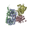
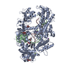
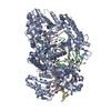
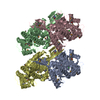
| Deposited unit | 
| ||||||||
|---|---|---|---|---|---|---|---|---|---|
| 1 |
| ||||||||
| Unit cell |
|
-Components
| #1: Protein | Mass: 17817.660 Da / Num. of mol.: 10 / Fragment: Carbohydrate recognition domain Source method: isolated from a genetically manipulated source Source: (gene. exp.) Homo sapiens (human) / Production host:  #2: Polysaccharide | 2-acetamido-2-deoxy-beta-D-glucopyranose-(1-2)-alpha-D-mannopyranose-(1-3)-[2-acetamido-2-deoxy- ...2-acetamido-2-deoxy-beta-D-glucopyranose-(1-2)-alpha-D-mannopyranose-(1-3)-[2-acetamido-2-deoxy-beta-D-glucopyranose-(1-2)-alpha-D-mannopyranose-(1-6)]alpha-D-mannopyranose Source method: isolated from a genetically manipulated source #3: Chemical | ChemComp-CA /   Mass: 40.078 Da / Num. of mol.: 30 / Source method: obtained synthetically / Formula: Ca Mass: 40.078 Da / Num. of mol.: 30 / Source method: obtained synthetically / Formula: Ca#4: Water | ChemComp-HOH / |  Mass: 18.015 Da / Num. of mol.: 252 / Source method: isolated from a natural source / Formula: H2O Mass: 18.015 Da / Num. of mol.: 252 / Source method: isolated from a natural source / Formula: H2OHas protein modification | Y | Sequence details | This alanine residue was inserted after the initiator methionine residue at the N-terminus to ...This alanine residue was inserted after the initiator methionine residue at the N-terminus to ensure that the methionine would be efficiently removed. This strategy ensures that the polypeptide has a free N-terminus, allowing Edman degradation to confirm the identity and integrity of the bacterially expressed protein. | |
|---|
-Experimental details
-Experiment
| Experiment | Method: X-RAY DIFFRACTION / Number of used crystals: 1 |
|---|
- Sample preparation
| Crystal | Density Matthews: 2.51 Å3/Da / Density % sol: 50.93 % | ||||||||||||||||||||||||||||||||||||||||||
|---|---|---|---|---|---|---|---|---|---|---|---|---|---|---|---|---|---|---|---|---|---|---|---|---|---|---|---|---|---|---|---|---|---|---|---|---|---|---|---|---|---|---|---|
| Crystal grow | Temperature: 294 K / Method: vapor diffusion, hanging drop / pH: 6.5 Details: 18% PEG 8000, 0.1M Na-cacodilate, 200mM Ca Ac2. Protein solution containing 5mM CaCl2 and 5 mM oligosaccharide (Dextra M592)., pH 6.5, VAPOR DIFFUSION, HANGING DROP, temperature 294K | ||||||||||||||||||||||||||||||||||||||||||
| Crystal grow | *PLUS Temperature: 21 ℃ | ||||||||||||||||||||||||||||||||||||||||||
| Components of the solutions | *PLUS
|
-Data collection
| Diffraction | Mean temperature: 100 K |
|---|---|
| Diffraction source | Source: SYNCHROTRON / Site: SSRL  / Beamline: BL11-1 / Wavelength: 0.9646 Å / Beamline: BL11-1 / Wavelength: 0.9646 Å |
| Detector | Type: ADSC QUANTUM 4 / Detector: CCD / Date: Apr 9, 2001 |
| Radiation | Protocol: SINGLE WAVELENGTH / Monochromatic (M) / Laue (L): M / Scattering type: x-ray |
| Radiation wavelength | Wavelength: 0.9646 Å / Relative weight: 1 |
| Reflection | Resolution: 2.5→50 Å / Num. all: 60699 / Num. obs: 59357 / % possible obs: 97.8 % / Observed criterion σ(F): 0 / Observed criterion σ(I): -3 / Biso Wilson estimate: 43 Å2 / Rmerge(I) obs: 0.053 |
| Reflection shell | Resolution: 2.5→2.59 Å / Rmerge(I) obs: 0.266 / % possible all: 99.3 |
| Reflection | *PLUS Lowest resolution: 50 Å / Num. measured all: 250720 |
| Reflection shell | *PLUS Highest resolution: 2.5 Å / % possible obs: 99.3 % |
- Processing
| Software |
| ||||||||||||||||||||||||||||||||||||||||||||||||||||||||||||||||||||||||||||||||
|---|---|---|---|---|---|---|---|---|---|---|---|---|---|---|---|---|---|---|---|---|---|---|---|---|---|---|---|---|---|---|---|---|---|---|---|---|---|---|---|---|---|---|---|---|---|---|---|---|---|---|---|---|---|---|---|---|---|---|---|---|---|---|---|---|---|---|---|---|---|---|---|---|---|---|---|---|---|---|---|---|---|
| Refinement | Method to determine structure: MOLECULAR REPLACEMENT / Resolution: 2.5→26.68 Å / Rfactor Rfree error: 0.004 / Data cutoff high absF: 2477698.65 / Data cutoff low absF: 0 / Isotropic thermal model: RESTRAINED / Cross valid method: THROUGHOUT / σ(F): 0
| ||||||||||||||||||||||||||||||||||||||||||||||||||||||||||||||||||||||||||||||||
| Solvent computation | Solvent model: FLAT MODEL / Bsol: 34.154 Å2 / ksol: 0.320351 e/Å3 | ||||||||||||||||||||||||||||||||||||||||||||||||||||||||||||||||||||||||||||||||
| Displacement parameters | Biso mean: 49.1 Å2
| ||||||||||||||||||||||||||||||||||||||||||||||||||||||||||||||||||||||||||||||||
| Refine analyze |
| ||||||||||||||||||||||||||||||||||||||||||||||||||||||||||||||||||||||||||||||||
| Refinement step | Cycle: LAST / Resolution: 2.5→26.68 Å
| ||||||||||||||||||||||||||||||||||||||||||||||||||||||||||||||||||||||||||||||||
| Refine LS restraints |
| ||||||||||||||||||||||||||||||||||||||||||||||||||||||||||||||||||||||||||||||||
| LS refinement shell | Resolution: 2.5→2.66 Å / Rfactor Rfree error: 0.013 / Total num. of bins used: 6
| ||||||||||||||||||||||||||||||||||||||||||||||||||||||||||||||||||||||||||||||||
| Xplor file |
| ||||||||||||||||||||||||||||||||||||||||||||||||||||||||||||||||||||||||||||||||
| Software | *PLUS Name: CNS / Version: 1 / Classification: refinement | ||||||||||||||||||||||||||||||||||||||||||||||||||||||||||||||||||||||||||||||||
| Refinement | *PLUS σ(F): 0 / % reflection Rfree: 6.1 % / Rfactor obs: 0.214 | ||||||||||||||||||||||||||||||||||||||||||||||||||||||||||||||||||||||||||||||||
| Solvent computation | *PLUS | ||||||||||||||||||||||||||||||||||||||||||||||||||||||||||||||||||||||||||||||||
| Displacement parameters | *PLUS Biso mean: 49.1 Å2 | ||||||||||||||||||||||||||||||||||||||||||||||||||||||||||||||||||||||||||||||||
| Refine LS restraints | *PLUS
| ||||||||||||||||||||||||||||||||||||||||||||||||||||||||||||||||||||||||||||||||
| LS refinement shell | *PLUS Rfactor Rfree: 0.328 / % reflection Rfree: 7.2 % / Rfactor Rwork: 0.291 |
