 Movie
Movie Controller

[English] 日本語
 Yorodumi
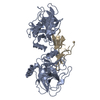
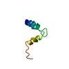
- PDB-1k9b: Crystal structure of the bifunctional soybean Bowman-Birk inhibit... -
+ Open data

- Basic information
| Entry | Database: PDB / ID: 1k9b | ||||||
|---|---|---|---|---|---|---|---|
| Title | Crystal structure of the bifunctional soybean Bowman-Birk inhibitor at 0.28 nm resolution. Structural peculiarities in a folded protein conformation | ||||||
 Components Components | BOWMAN-BIRK TYPE PROTEINASE INHIBITOR | ||||||
 Keywords Keywords | HYDROLASE INHIBITOR / TRIPPLE-STRANDED BETA HAIRPIN / DOUBLE-HEADED | ||||||
| Function / homology |  Function and homology information Function and homology information | ||||||
| Biological species |  | ||||||
| Method |  X-RAY DIFFRACTION / SYNCHROTRON / MOLECULAR REPLACEMENT / Resolution: 2.8 Å X-RAY DIFFRACTION / SYNCHROTRON / MOLECULAR REPLACEMENT / Resolution: 2.8 Å | ||||||
 Authors Authors | Voss, R.H. / Ermler, U. / Essen, L.O. / Wenzl, G. / Kim, Y.M. / Flecker, P. | ||||||
 Citation Citation | Journal: Eur.J.Biochem. / Year: 1996 Title: Crystal structure of the bifunctional soybean Bowman-Birk inhibitor at 0.28-nm resolution. Structural peculiarities in a folded protein conformation. Authors: Voss, R.H. / Ermler, U. / Essen, L.O. / Wenzl, G. / Kim, Y.M. / Flecker, P. #1: Journal: J.Mol.Biol. / Year: 2000Title: Crystal Structure of Cancer Chemopreventive Bowman-Birk Inhibitor in Ternary Complex with Bovine Trypsin at 2.3 Ao Resolution. Structural Basis of Janus-faced Serine Protease Inhibitor Specificity Authors: Koepke, J. / Ermler, U. / Warkentin, E. / Wenzl, G. / Flecker, P. #2: Journal: Eur.J.Biochem. / Year: 1987Title: Chemical synthesis, molecular cloning and expression of gene coding for a Bowman-Birk-type proteinase inhibitor Authors: Flecker, P. #3: Journal: Eur.J.Biochem. / Year: 1998Title: Mutational analysis of disulfide bonds in the trypsin-reactive subdomain of a Bowman-Birk-type inhibitor of trypsin and chymotrypsin. Cooperative versus autonomous refolding of subdomains Authors: Philipp, S. / Kim, Y.M. / Duerr, I. / Wenzl, G. / Vogt, M. / Flecker, P. #4: Journal: Eur.J.Biochem. / Year: 1995Title: Template-directed protein folding into a metastable state of increased activity Authors: Flecker, P. #5: Journal: FEBS Lett. / Year: 1989Title: A new and general procedure for refolding mutant Bowman-Birk-type proteinase inhibitors on trypsin-Sepharose as a matrix with complementary structure. Authors: Flecker, P. | ||||||
| History |
|
- Structure visualization
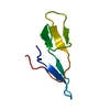
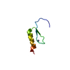
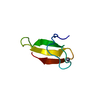
| Structure viewer | Molecule: MolmilJmol/JSmol |
|---|
- Downloads & links
-Download
| PDBx/mmCIF format | 1k9b.cif.gz | 25.4 KB | Display | PDBx/mmCIF format |
|---|---|---|---|---|
| PDB format | pdb1k9b.ent.gz | 16.4 KB | Display | PDB format |
| PDBx/mmJSON format | 1k9b.json.gz | Tree view | PDBx/mmJSON format | |
| Others |  Other downloads Other downloads |
-Validation report
| Arichive directory | https://data.pdbj.org/pub/pdb/validation_reports/k9/1k9bftp://data.pdbj.org/pub/pdb/validation_reports/k9/1k9b | HTTPS FTP |
|---|
-Related structure data
| Related structure data |  1pi2S S: Starting model for refinement |
|---|---|
| Similar structure data |


-Links
 PDBj
PDBj- Assembly
| Deposited unit | 
| ||||||||||
|---|---|---|---|---|---|---|---|---|---|---|---|
| 1 |
| ||||||||||
| Unit cell |
|
-Components
| #1: Protein | Mass: 6431.538 Da / Num. of mol.: 1 / Source method: isolated from a natural source / Details: free form / Source: (natural) |
|---|---|
| #2: Water | ChemComp-HOH /  Mass: 18.015 Da / Num. of mol.: 17 / Source method: isolated from a natural source / Formula: H2O Mass: 18.015 Da / Num. of mol.: 17 / Source method: isolated from a natural source / Formula: H2O |
| Has protein modification | Y |
-Experimental details
-Experiment
| Experiment | Method: X-RAY DIFFRACTION / Number of used crystals: 1 |
|---|
- Sample preparation
| Crystal | Density Matthews: 4.13 Å3/Da / Density % sol: 70.23 % |
|---|---|
| Crystal grow | Temperature: 289 K / Method: vapor diffusion, sitting drop / pH: 7.6 Details: PEG 4000, ammonium sulfate, pH 7.6, VAPOR DIFFUSION, SITTING DROP at 289K |
-Data collection
| Diffraction | Mean temperature: 277 K |
|---|---|
| Diffraction source | Source: SYNCHROTRON / Site: Photon Factory  / Beamline: BL-6A / Wavelength: 1.04 Å / Beamline: BL-6A / Wavelength: 1.04 Å |
| Detector | Type: WEISSENBERG / Detector: DIFFRACTOMETER / Date: Apr 5, 1994 |
| Radiation | Protocol: SINGLE WAVELENGTH / Monochromatic (M) / Laue (L): M / Scattering type: x-ray |
| Radiation wavelength | Wavelength: 1.04 Å / Relative weight: 1 |
| Reflection | Resolution: 2.5→60.9 Å / Num. all: 3891 / Num. obs: 26756 / % possible obs: 96.3 % / Observed criterion σ(I): -3 / Redundancy: 6.88 % / Rmerge(I) obs: 0.058 / Rsym value: 0.058 |
| Reflection shell | Resolution: 2.8→3 Å / Rmerge(I) obs: 0.3 / % possible all: 70 |
- Processing
| Software |
| |||||||||||||||||||||||||||
|---|---|---|---|---|---|---|---|---|---|---|---|---|---|---|---|---|---|---|---|---|---|---|---|---|---|---|---|---|
| Refinement | Method to determine structure: MOLECULAR REPLACEMENT Starting model: PDB ENTRY 1PI2 Resolution: 2.8→8 Å / Cross valid method: THROUGHOUT / σ(F): 2 / Stereochemistry target values: Engh & Huber
| |||||||||||||||||||||||||||
| Displacement parameters | Biso mean: 28.7 Å2 | |||||||||||||||||||||||||||
| Refine analyze | Luzzati coordinate error obs: 0.035 Å | |||||||||||||||||||||||||||
| Refinement step | Cycle: LAST / Resolution: 2.8→8 Å
| |||||||||||||||||||||||||||
| Refine LS restraints |
|
