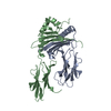
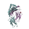
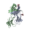
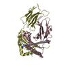
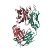
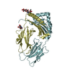
 Movie
Movie Controller


+ Open data

- Basic information
| Entry | Database: PDB / ID: 1k8i | ||||||
|---|---|---|---|---|---|---|---|
| Title | CRYSTAL STRUCTURE OF MOUSE H2-DM | ||||||
 Components Components |
| ||||||
 Keywords Keywords | IMMUNE SYSTEM / MHC | ||||||
| Function / homology |  Function and homology information Function and homology informationMHC class II protein complex assembly / positive regulation of humoral immune response / positive regulation of germinal center formation / positive regulation of T cell activation via T cell receptor contact with antigen bound to MHC molecule on antigen presenting cell / MHC class II antigen presentation / positive thymic T cell selection / positive regulation of T cell differentiation / antigen processing and presentation / immunoglobulin mediated immune response / inner ear development ...MHC class II protein complex assembly / positive regulation of humoral immune response / positive regulation of germinal center formation / positive regulation of T cell activation via T cell receptor contact with antigen bound to MHC molecule on antigen presenting cell / MHC class II antigen presentation / positive thymic T cell selection / positive regulation of T cell differentiation / antigen processing and presentation / immunoglobulin mediated immune response / inner ear development / multivesicular body / positive regulation of T cell proliferation / peptide antigen assembly with MHC class II protein complex / MHC class II protein complex / antigen processing and presentation of exogenous peptide antigen via MHC class II / positive regulation of immune response / peptide antigen binding / positive regulation of T cell activation / MHC class II protein complex binding / late endosome / late endosome membrane / protein transport / protein folding / protein-containing complex assembly / adaptive immune response / lysosome / endosome membrane / lysosomal membrane / cell surface / membrane Similarity search - Function | ||||||
| Biological species |  | ||||||
| Method |  X-RAY DIFFRACTION / molecular replacement,MIR / Resolution: 3.1 Å X-RAY DIFFRACTION / molecular replacement,MIR / Resolution: 3.1 Å | ||||||
 Authors Authors | Fremont, D.H. / Crawford, F. / Marrack, P. / Hendrickson, W. / Kappler, J. | ||||||
 Citation Citation | Journal: Immunity / Year: 1998 Title: Crystal structure of mouse H2-M. Authors: Fremont, D.H. / Crawford, F. / Marrack, P. / Hendrickson, W.A. / Kappler, J. | ||||||
| History |
|
- Structure visualization
| Structure viewer | Molecule: MolmilJmol/JSmol |
|---|
- Downloads & links
-Download
| PDBx/mmCIF format | 1k8i.cif.gz | 85.7 KB | Display | PDBx/mmCIF format |
|---|---|---|---|---|
| PDB format | pdb1k8i.ent.gz | 65.2 KB | Display | PDB format |
| PDBx/mmJSON format | 1k8i.json.gz | Tree view | PDBx/mmJSON format | |
| Others |  Other downloads Other downloads |
-Validation report
| Arichive directory | https://data.pdbj.org/pub/pdb/validation_reports/k8/1k8iftp://data.pdbj.org/pub/pdb/validation_reports/k8/1k8i | HTTPS FTP |
|---|
-Related structure data
| Similar structure data |
|---|


-Links
 PDBj
PDBj


- Assembly
| Deposited unit | 
| ||||||||
|---|---|---|---|---|---|---|---|---|---|
| 1 |
| ||||||||
| Unit cell |
|
-Components
| #1: Protein | Mass: 21466.945 Da / Num. of mol.: 1 Source method: isolated from a genetically manipulated source Source: (gene. exp.)  Trichoplusia ni (cabbage looper) / References: UniProt: P28078 Trichoplusia ni (cabbage looper) / References: UniProt: P28078 | ||
|---|---|---|---|
| #2: Protein | Mass: 21329.883 Da / Num. of mol.: 1 Source method: isolated from a genetically manipulated source Source: (gene. exp.) Trichoplusia ni (cabbage looper) / References: UniProt: Q31099 | ||
| #3: Sugar |   Type: D-saccharide, beta linking / Mass: 221.208 Da / Num. of mol.: 2 Type: D-saccharide, beta linking / Mass: 221.208 Da / Num. of mol.: 2Source method: isolated from a genetically manipulated source Formula: C8H15NO6 Has protein modification | Y | |
-Experimental details
-Experiment
| Experiment | Method: X-RAY DIFFRACTION / Number of used crystals: 4 |
|---|
- Sample preparation
| Crystal | Density Matthews: 4.57 Å3/Da / Density % sol: 73.09 % | ||||||||||||||||||||||||||||||||||||||||||
|---|---|---|---|---|---|---|---|---|---|---|---|---|---|---|---|---|---|---|---|---|---|---|---|---|---|---|---|---|---|---|---|---|---|---|---|---|---|---|---|---|---|---|---|
| Crystal grow | Temperature: 293 K / Method: vapor diffusion, hanging drop / pH: 5.5 Details: 0.1 M lithium sulfate, 0.1M MES, pH 5.5, VAPOR DIFFUSION, HANGING DROP, temperature 293K | ||||||||||||||||||||||||||||||||||||||||||
| Crystal grow | *PLUS Temperature: 20 ℃ / Details: used microseeding | ||||||||||||||||||||||||||||||||||||||||||
| Components of the solutions | *PLUS
|
-Data collection
| Diffraction | Mean temperature: 298 K |
|---|---|
| Diffraction source | Source: ROTATING ANODE / Type: RIGAKU / Wavelength: 1.5 Å |
| Detector | Type: RIGAKU RAXIS IV / Detector: IMAGE PLATE / Date: Apr 1, 1998 / Details: mirrors |
| Radiation | Monochromator: Yale mirrors / Protocol: SINGLE WAVELENGTH / Monochromatic (M) / Laue (L): M / Scattering type: x-ray |
| Radiation wavelength | Wavelength: 1.5 Å / Relative weight: 1 |
| Reflection | Resolution: 3.1→20 Å / Num. all: 13888 / Num. obs: 13711 / % possible obs: 98.1 % / Observed criterion σ(F): 1 / Observed criterion σ(I): -3 / Redundancy: 4.56 % / Biso Wilson estimate: 74.5 Å2 / Rsym value: 0.091 / Net I/σ(I): 16.2 |
| Reflection shell | Resolution: 3.1→3.2 Å / Mean I/σ(I) obs: 1.71 / Num. unique all: 1303 / Rsym value: 0.482 / % possible all: 95.2 |
| Reflection | *PLUS Lowest resolution: 20 Å / Num. obs: 13888 / Num. measured all: 63314 / Rmerge(I) obs: 0.091 |
| Reflection shell | *PLUS % possible obs: 95.2 % / Rmerge(I) obs: 0.482 |
- Processing
| Software |
| |||||||||||||||||||||||||
|---|---|---|---|---|---|---|---|---|---|---|---|---|---|---|---|---|---|---|---|---|---|---|---|---|---|---|
| Refinement | Method to determine structure: molecular replacement,MIR / Resolution: 3.1→12 Å / Rfactor Rfree error: 0.01 / Data cutoff high absF: 100000 / Data cutoff low absF: 0.01 / Isotropic thermal model: RESTRAINED / Cross valid method: THROUGHOUT / σ(F): 0
| |||||||||||||||||||||||||
| Displacement parameters | Biso mean: 64.1 Å2
| |||||||||||||||||||||||||
| Refine analyze |
| |||||||||||||||||||||||||
| Refinement step | Cycle: LAST / Resolution: 3.1→12 Å
| |||||||||||||||||||||||||
| Refine LS restraints |
| |||||||||||||||||||||||||
| LS refinement shell | Resolution: 3.1→3.21 Å / Rfactor Rfree error: 0.038 / Total num. of bins used: 10
| |||||||||||||||||||||||||
| Xplor file |
| |||||||||||||||||||||||||
| Refinement | *PLUS Highest resolution: 3.1 Å / Lowest resolution: 12 Å / % reflection Rfree: 5 % | |||||||||||||||||||||||||
| Solvent computation | *PLUS | |||||||||||||||||||||||||
| Displacement parameters | *PLUS | |||||||||||||||||||||||||
| Refine LS restraints | *PLUS
| |||||||||||||||||||||||||
| LS refinement shell | *PLUS Highest resolution: 3.1 Å / Lowest resolution: 3.2 Å / Rfactor Rfree: 0.334 / Rfactor Rwork: 0.345 / Num. reflection obs: 1303 / Rfactor obs: 0.345 |
