 Movie
Movie Controller


+ Open data

- Basic information
| Entry | Database: PDB / ID: 1k7h | ||||||
|---|---|---|---|---|---|---|---|
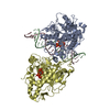
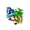
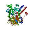
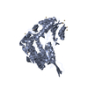
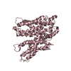
| Title | CRYSTAL STRUCTURE OF SHRIMP ALKALINE PHOSPHATASE | ||||||
 Components Components | ALKALINE PHOSPHATASE | ||||||
 Keywords Keywords | HYDROLASE / TRANSFERASE / PHOSPHOMONOESTER / EXTENDED BETA SHEET / ZINC TRIAD / METAL TRIAD | ||||||
| Function / homology |  Function and homology information Function and homology informationalkaline phosphatase / alkaline phosphatase activity / side of membrane / metal ion binding / plasma membrane Similarity search - Function | ||||||
| Biological species |  Pandalus borealis (northern shrimp) Pandalus borealis (northern shrimp) | ||||||
| Method |  X-RAY DIFFRACTION / SYNCHROTRON / MAD / Resolution: 1.92 Å X-RAY DIFFRACTION / SYNCHROTRON / MAD / Resolution: 1.92 Å | ||||||
 Authors Authors | De Backer, M.E. / Mc Sweeney, S. / Rasmussen, H.B. / Riise, B.W. / Lindley, P. / Hough, E. | ||||||
 Citation Citation | Journal: J.Mol.Biol. / Year: 2002 Title: The 1.9 A Crystal Structure of Heat-Labile Shrimp Alkaline Phosphatase Authors: De Backer, M.E. / Mc Sweeney, S. / Rasmussen, H.B. / Riise, B.W. / Lindley, P. / Hough, E. #1: Journal: J.Biol.Chem. / Year: 2001Title: Crystal Structure of Alkaline Phosphatase from Huma at 1.8 A Resolution. Implication for a Substrate Specificity Authors: Le Du, M.H. / Stigbrand, T. / Taussig, M.J. / Menez, A. / Stura, E.A. #2: Journal: J.Mol.Biol. / Year: 1991Title: Reaction Mechanism of Alkaline Phosphatase Based on Crystal Structures. Two-Metal Ion Catalysis Authors: Kim, E.E. / Wyckoff, H.W. | ||||||
| History |
|
- Structure visualization
| Structure viewer | Molecule: MolmilJmol/JSmol |
|---|
- Downloads & links
-Download
| PDBx/mmCIF format | 1k7h.cif.gz | 209.7 KB | Display | PDBx/mmCIF format |
|---|---|---|---|---|
| PDB format | pdb1k7h.ent.gz | 166.6 KB | Display | PDB format |
| PDBx/mmJSON format | 1k7h.json.gz | Tree view | PDBx/mmJSON format | |
| Others |  Other downloads Other downloads |
-Validation report
| Arichive directory | https://data.pdbj.org/pub/pdb/validation_reports/k7/1k7hftp://data.pdbj.org/pub/pdb/validation_reports/k7/1k7h | HTTPS FTP |
|---|
-Related structure data
| Similar structure data |
|---|


-Links
 PDBj
PDBj
- Assembly
| Deposited unit | 
| ||||||||
|---|---|---|---|---|---|---|---|---|---|
| 1 | 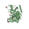
| ||||||||
| 2 | 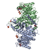
| ||||||||
| Unit cell |
|
-Components
-Protein / Sugars , 2 types, 4 molecules AB
| #1: Protein | Mass: 53032.336 Da / Num. of mol.: 2 / Source method: isolated from a natural source / Source: (natural) Pandalus borealis (northern shrimp) / References: UniProt: Q9BHT8, alkaline phosphatase#2: Sugar |  Type: D-saccharide, beta linking / Mass: 221.208 Da / Num. of mol.: 2 Type: D-saccharide, beta linking / Mass: 221.208 Da / Num. of mol.: 2Source method: isolated from a genetically manipulated source Formula: C8H15NO6 |
|---|
-Non-polymers , 4 types, 563 molecules 


| #3: Chemical | ChemComp-ZN /  Mass: 65.409 Da / Num. of mol.: 6 / Source method: obtained synthetically / Formula: Zn Mass: 65.409 Da / Num. of mol.: 6 / Source method: obtained synthetically / Formula: Zn#4: Chemical | ChemComp-SO4 /  Mass: 96.063 Da / Num. of mol.: 6 / Source method: obtained synthetically / Formula: SO4 Mass: 96.063 Da / Num. of mol.: 6 / Source method: obtained synthetically / Formula: SO4#5: Chemical |  Mass: 116.072 Da / Num. of mol.: 3 / Source method: obtained synthetically / Formula: C4H4O4 Mass: 116.072 Da / Num. of mol.: 3 / Source method: obtained synthetically / Formula: C4H4O4#6: Water | ChemComp-HOH / | Mass: 18.015 Da / Num. of mol.: 548 / Source method: isolated from a natural source / Formula: H2O |
|---|
-Details
| Has protein modification | Y |
|---|
-Experimental details
-Experiment
| Experiment | Method: X-RAY DIFFRACTION / Number of used crystals: 1 |
|---|
- Sample preparation
| Crystal | Density Matthews: 2.92 Å3/Da / Density % sol: 47 % | ||||||||||||||||||||||||||||||||||||||||||
|---|---|---|---|---|---|---|---|---|---|---|---|---|---|---|---|---|---|---|---|---|---|---|---|---|---|---|---|---|---|---|---|---|---|---|---|---|---|---|---|---|---|---|---|
| Crystal grow | pH: 5.6 / Details: pH 5.60 | ||||||||||||||||||||||||||||||||||||||||||
| Crystal grow | *PLUS pH: 5.6 / Method: vapor diffusion, hanging drop | ||||||||||||||||||||||||||||||||||||||||||
| Components of the solutions | *PLUS
|
-Data collection
| Diffraction | Mean temperature: 100 K | |||||||||||||||
|---|---|---|---|---|---|---|---|---|---|---|---|---|---|---|---|---|
| Diffraction source | Source: SYNCHROTRON / Site: ESRF  / Beamline: ID14-4 / Wavelength: 0.9310,1.2820,1.2825,0.933 / Beamline: ID14-4 / Wavelength: 0.9310,1.2820,1.2825,0.933 | |||||||||||||||
| Detector | Type: ADSC QUANTUM 4r / Detector: CCD / Date: Jun 1, 2000 / Details: TOROIDAL MIRROR | |||||||||||||||
| Radiation | Monochromator: DIAMOND(111) DIAMOND(111) / Protocol: SINGLE WAVELENGTH / Monochromatic (M) / Laue (L): M / Scattering type: x-ray | |||||||||||||||
| Radiation wavelength |
| |||||||||||||||
| Reflection | Resolution: 1.85→99 Å / Num. obs: 95547 / % possible obs: 96.8 % / Observed criterion σ(I): 0 / Redundancy: 2.6 % / Biso Wilson estimate: 24.2 Å2 / Rmerge(I) obs: 0.076 / Rsym value: 0.176 / Net I/σ(I): 11.8 | |||||||||||||||
| Reflection shell | Resolution: 1.92→1.99 Å / Redundancy: 2.5 % / Rmerge(I) obs: 0.465 / Mean I/σ(I) obs: 2.2 / Rsym value: 0.597 / % possible all: 99.2 | |||||||||||||||
| Reflection | *PLUS Num. obs: 103370 / Redundancy: 2.6 % / Num. measured all: 1131014 | |||||||||||||||
| Reflection shell | *PLUS % possible obs: 99.2 % / Mean I/σ(I) obs: 2.2 |
- Processing
| Software |
| |||||||||||||||||||||||||||||||||||||||||||||||||||||||||||||||||||||||||||||||||||||||||||||||||||||||||||||||||||||||||||||||||||||||
|---|---|---|---|---|---|---|---|---|---|---|---|---|---|---|---|---|---|---|---|---|---|---|---|---|---|---|---|---|---|---|---|---|---|---|---|---|---|---|---|---|---|---|---|---|---|---|---|---|---|---|---|---|---|---|---|---|---|---|---|---|---|---|---|---|---|---|---|---|---|---|---|---|---|---|---|---|---|---|---|---|---|---|---|---|---|---|---|---|---|---|---|---|---|---|---|---|---|---|---|---|---|---|---|---|---|---|---|---|---|---|---|---|---|---|---|---|---|---|---|---|---|---|---|---|---|---|---|---|---|---|---|---|---|---|---|---|
| Refinement | Method to determine structure: MAD / Resolution: 1.92→119.5 Å / Cor.coef. Fo:Fc: 0.95 / Cor.coef. Fo:Fc free: 0.942 / SU B: 4.79 / SU ML: 0.14 / Cross valid method: THROUGHOUT / σ(F): 0 / ESU R: 0.149 / ESU R Free: 0.136 / Stereochemistry target values: MAXIMUM LIKELIHOOD / Details: HYDROGENS HAVE BEEN ADDED IN THE RIDING POSITIONS
| |||||||||||||||||||||||||||||||||||||||||||||||||||||||||||||||||||||||||||||||||||||||||||||||||||||||||||||||||||||||||||||||||||||||
| Solvent computation | Ion probe radii: 0.8 Å / Shrinkage radii: 0.8 Å / VDW probe radii: 1.4 Å / Solvent model: BABINET MODEL WITH MASK | |||||||||||||||||||||||||||||||||||||||||||||||||||||||||||||||||||||||||||||||||||||||||||||||||||||||||||||||||||||||||||||||||||||||
| Displacement parameters | Biso mean: 25.99 Å2
| |||||||||||||||||||||||||||||||||||||||||||||||||||||||||||||||||||||||||||||||||||||||||||||||||||||||||||||||||||||||||||||||||||||||
| Refinement step | Cycle: LAST / Resolution: 1.92→119.5 Å
| |||||||||||||||||||||||||||||||||||||||||||||||||||||||||||||||||||||||||||||||||||||||||||||||||||||||||||||||||||||||||||||||||||||||
| Refine LS restraints |
| |||||||||||||||||||||||||||||||||||||||||||||||||||||||||||||||||||||||||||||||||||||||||||||||||||||||||||||||||||||||||||||||||||||||
| LS refinement shell | Resolution: 1.92→1.97 Å / Total num. of bins used: 20 /
| |||||||||||||||||||||||||||||||||||||||||||||||||||||||||||||||||||||||||||||||||||||||||||||||||||||||||||||||||||||||||||||||||||||||
| Refinement | *PLUS % reflection Rfree: 4.9 % | |||||||||||||||||||||||||||||||||||||||||||||||||||||||||||||||||||||||||||||||||||||||||||||||||||||||||||||||||||||||||||||||||||||||
| Solvent computation | *PLUS | |||||||||||||||||||||||||||||||||||||||||||||||||||||||||||||||||||||||||||||||||||||||||||||||||||||||||||||||||||||||||||||||||||||||
| Displacement parameters | *PLUS |
