 Movie
Movie Controller

[English] 日本語
 Yorodumi
- PDB-1k75: The L-histidinol dehydrogenase (hisD) structure implicates domain... -
+ Open data

- Basic information
| Entry | Database: PDB / ID: 1k75 | ||||||
|---|---|---|---|---|---|---|---|
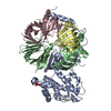
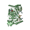
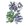
| Title | The L-histidinol dehydrogenase (hisD) structure implicates domain swapping and gene duplication. | ||||||
 Components Components | L-histidinol dehydrogenase | ||||||
 Keywords Keywords | OXIDOREDUCTASE / L-histidinol dehydrogenase / homodimer / Rossmann fold / 4 domains / hisD / L-histidine biosynthesis / NAD cofactor / Montreal-Kingston Bacterial Structural Genomics Initiative / BSGI / Structural Genomics | ||||||
| Function / homology |  Function and homology information Function and homology informationhistidinol dehydrogenase / histidinol dehydrogenase activity / L-histidine biosynthetic process / NAD binding / manganese ion binding / zinc ion binding / metal ion binding / cytosol / cytoplasm Similarity search - Function | ||||||
| Biological species |  | ||||||
| Method |  X-RAY DIFFRACTION / SYNCHROTRON / MAD / Resolution: 1.75 Å X-RAY DIFFRACTION / SYNCHROTRON / MAD / Resolution: 1.75 Å | ||||||
 Authors Authors | Barbosa, J.A.R.G. / Sivaraman, J. / Li, Y. / Larocque, R. / Matte, A. / Schrag, J. / Cygler, M. / Montreal-Kingston Bacterial Structural Genomics Initiative (BSGI) | ||||||
 Citation Citation | Journal: Proc.Natl.Acad.Sci.USA / Year: 2002 Title: Mechanism of action and NAD+-binding mode revealed by the crystal structure of L-histidinol dehydrogenase. Authors: Barbosa, J.A.R.G. / Sivaraman, J. / Li, Y. / Larocque, R. / Matte, A. / Schrag, J.D. / Cygler, M. | ||||||
| History |
|
- Structure visualization
| Structure viewer | Molecule: MolmilJmol/JSmol |
|---|
- Downloads & links
-Download
| PDBx/mmCIF format | 1k75.cif.gz | 195.9 KB | Display | PDBx/mmCIF format |
|---|---|---|---|---|
| PDB format | pdb1k75.ent.gz | 153.6 KB | Display | PDB format |
| PDBx/mmJSON format | 1k75.json.gz | Tree view | PDBx/mmJSON format | |
| Others |  Other downloads Other downloads |
-Validation report
| Arichive directory | https://data.pdbj.org/pub/pdb/validation_reports/k7/1k75ftp://data.pdbj.org/pub/pdb/validation_reports/k7/1k75 | HTTPS FTP |
|---|
-Related structure data
| Related structure data |  1kaeC  1kahC  1karC C: citing same article ( |
|---|---|
| Similar structure data | |
| Other databases |


-Links
 PDBj
PDBj

- Assembly
| Deposited unit | 
| ||||||||
|---|---|---|---|---|---|---|---|---|---|
| 1 |
| ||||||||
| Unit cell |
| ||||||||
| Details | The biological (functional) hisD is the homodimer in the assymetric unit. No symmetry operations are needed. |
-Components
| #1: Protein | Mass: 46434.520 Da / Num. of mol.: 2 / Fragment: Se-Met derived dimer Source method: isolated from a genetically manipulated source Source: (gene. exp.) #2: Chemical | ChemComp-SO4 /   Mass: 96.063 Da / Num. of mol.: 4 / Source method: obtained synthetically / Formula: SO4 Mass: 96.063 Da / Num. of mol.: 4 / Source method: obtained synthetically / Formula: SO4#3: Chemical | ChemComp-GOL / |   Mass: 92.094 Da / Num. of mol.: 1 / Source method: obtained synthetically / Formula: C3H8O3 Mass: 92.094 Da / Num. of mol.: 1 / Source method: obtained synthetically / Formula: C3H8O3#4: Water | ChemComp-HOH / |  Mass: 18.015 Da / Num. of mol.: 924 / Source method: isolated from a natural source / Formula: H2O Mass: 18.015 Da / Num. of mol.: 924 / Source method: isolated from a natural source / Formula: H2OHas protein modification | Y | |
|---|
-Experimental details
-Experiment
| Experiment | Method: X-RAY DIFFRACTION / Number of used crystals: 1 |
|---|
- Sample preparation
| Crystal | Density Matthews: 2.47 Å3/Da / Density % sol: 50.26 % | ||||||||||||||||||||||||||||||||||||||||||||||||||||||||||||||||||||||
|---|---|---|---|---|---|---|---|---|---|---|---|---|---|---|---|---|---|---|---|---|---|---|---|---|---|---|---|---|---|---|---|---|---|---|---|---|---|---|---|---|---|---|---|---|---|---|---|---|---|---|---|---|---|---|---|---|---|---|---|---|---|---|---|---|---|---|---|---|---|---|---|
| Crystal grow | Temperature: 291 K / Method: vapor diffusion, hanging drop / pH: 7.5 Details: PEG 3350, glycerol, imidazole/malic acid buffer, ammonium sulfate, pH 7.5, VAPOR DIFFUSION, HANGING DROP, temperature 291K | ||||||||||||||||||||||||||||||||||||||||||||||||||||||||||||||||||||||
| Crystal grow | *PLUS Temperature: 18 ℃ | ||||||||||||||||||||||||||||||||||||||||||||||||||||||||||||||||||||||
| Components of the solutions | *PLUS
|
-Data collection
| Diffraction | Mean temperature: 100 K | ||||||||||||
|---|---|---|---|---|---|---|---|---|---|---|---|---|---|
| Diffraction source | Source: SYNCHROTRON / Site: NSLS  / Beamline: X8C / Wavelength: 0.97950,0.97934,0.97857 / Beamline: X8C / Wavelength: 0.97950,0.97934,0.97857 | ||||||||||||
| Detector | Type: ADSC QUANTUM 4 / Detector: CCD / Date: Jul 15, 2000 / Details: mirrors | ||||||||||||
| Radiation | Protocol: MAD / Monochromatic (M) / Laue (L): M / Scattering type: x-ray | ||||||||||||
| Radiation wavelength |
| ||||||||||||
| Reflection | Resolution: 1.75→40 Å / Num. all: 91930 / Num. obs: 91930 / % possible obs: 97.3 % / Observed criterion σ(F): 0 / Observed criterion σ(I): 0 / Redundancy: 4.4 % / Biso Wilson estimate: 19.7 Å2 / Rsym value: 0.06 / Net I/σ(I): 11.4 | ||||||||||||
| Reflection shell | Resolution: 1.75→1.78 Å / Redundancy: 2.7 % / Mean I/σ(I) obs: 2.2 / Num. unique all: 3675 / Rsym value: 0.276 / % possible all: 79.7 | ||||||||||||
| Reflection | *PLUS Num. obs: 91330 / Num. measured all: 402344 / Rmerge(I) obs: 0.06 | ||||||||||||
| Reflection shell | *PLUS % possible obs: 79.7 % / Rmerge(I) obs: 0.276 |
- Processing
| Software |
| |||||||||||||||||||||||||
|---|---|---|---|---|---|---|---|---|---|---|---|---|---|---|---|---|---|---|---|---|---|---|---|---|---|---|
| Refinement | Method to determine structure: MAD / Resolution: 1.75→40 Å / Isotropic thermal model: isotropic / Cross valid method: THROUGHOUT / σ(F): 0 / σ(I): 0 / Stereochemistry target values: Engh & Huber / Details: Used bulk solvent correction
| |||||||||||||||||||||||||
| Displacement parameters | Biso mean: 23.4 Å2
| |||||||||||||||||||||||||
| Refine analyze |
| |||||||||||||||||||||||||
| Refinement step | Cycle: LAST / Resolution: 1.75→40 Å
| |||||||||||||||||||||||||
| Refine LS restraints |
| |||||||||||||||||||||||||
| LS refinement shell | Resolution: 1.75→1.81 Å / Rfactor Rfree error: 0.019
| |||||||||||||||||||||||||
| Software | *PLUS Name: CNS / Version: 1 / Classification: refinement | |||||||||||||||||||||||||
| Refinement | *PLUS σ(F): 0 / Rfactor obs: 0.19 / Rfactor Rwork: 0.19 | |||||||||||||||||||||||||
| Solvent computation | *PLUS | |||||||||||||||||||||||||
| Displacement parameters | *PLUS | |||||||||||||||||||||||||
| Refine LS restraints | *PLUS
| |||||||||||||||||||||||||
| LS refinement shell | *PLUS Lowest resolution: 1.78 Å / Rfactor Rwork: 0.244 / Rfactor obs: 0.244 |
